

Blood Flow Mimicking Aneurysmal Wall Enhancement: A Diagnostic Pitfall of Vessel Wall MRI Using the Postcontrast 3D Turbo Spin-Echo MR Imaging Sequence
- PMID: 29599170
- PMCID: PMC7410621
- DOI: 10.3174/ajnr.A5616
Blood Flow Mimicking Aneurysmal Wall Enhancement: A Diagnostic Pitfall of Vessel Wall MRI Using the Postcontrast 3D Turbo Spin-Echo MR Imaging Sequence
Abstract
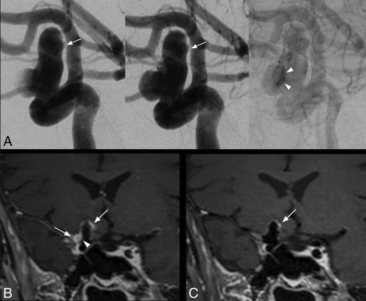
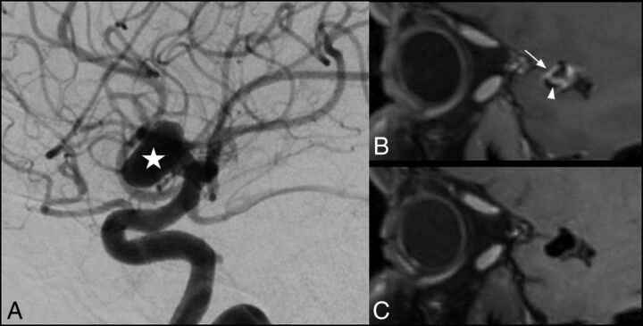
Our aim was to compare the detectability of aneurysmal wall enhancement in unruptured intracranial aneurysms between conventional and motion-sensitized driven equilibrium-prepared postcontrast 3D T1-weighted TSE sequences (sampling perfection with applicationoptimized contrasts by using different flip angle evolution, SPACE). Twenty-two patients with 30 unruptured intracranial aneurysms were scanned at 3T. Aneurysmal wall enhancement was more significantly detected using conventional compared with motion-sensitized driven equilibrium-prepared SPACE sequences (10/30 versus 2/30, P < .0001). Contrast-to-noise ratio measurements did not differ between conventional and motion-sensitized driven equilibrium-prepared sequences (P = .51). Flowing blood can mimic aneurysmal wall enhancement using conventional SPACE sequences with potential implications for patient care.
© 2018 by American Journal of Neuroradiology.
Figures
Comment in
-
Comment on "Blood Flow Mimicking Aneurysmal Wall Enhancement: A Diagnostic Pitfall of Vessel Wall MRI Using the Postcontrast 3D Turbo Spin-Echo MR Imaging Sequence".AJNR Am J Neuroradiol. 2018 Nov;39(11):E118. doi: 10.3174/ajnr.A5777. Epub 2018 Sep 6. AJNR Am J Neuroradiol. 2018. PMID: 30190255 Free PMC article. No abstract available.
-
Reply.AJNR Am J Neuroradiol. 2018 Nov;39(11):E119. doi: 10.3174/ajnr.A5789. Epub 2018 Sep 6. AJNR Am J Neuroradiol. 2018. PMID: 30190256 Free PMC article. No abstract available.
Similar articles
-
Wall Enhancement of the Intracranial Aneurysms Revealed by Magnetic Resonance Vessel Wall Imaging Using Three-Dimensional Turbo Spin-Echo Sequence with Motion-Sensitized Driven-Equilibrium: A Sign of Ruptured Aneurysm?Clin Neuroradiol. 2016 Sep;26(3):277-83. doi: 10.1007/s00062-014-0353-z. Epub 2014 Oct 21. Clin Neuroradiol. 2016. PMID: 25332151
-
Insufficient slow-flow suppression mimicking aneurysm wall enhancement in magnetic resonance vessel wall imaging: a phantom study.Neurosurg Focus. 2019 Jul 1;47(1):E19. doi: 10.3171/2019.4.FOCUS19235. Neurosurg Focus. 2019. PMID: 31261123
-
Visualizing wall enhancement over time in unruptured intracranial aneurysms using 3D vessel wall imaging.J Magn Reson Imaging. 2019 Jul;50(1):193-200. doi: 10.1002/jmri.26553. Epub 2018 Nov 3. J Magn Reson Imaging. 2019. PMID: 30390363
-
Conventional and high-resolution vessel wall MRI of intracranial aneurysms: current concepts and new horizons.J Neurosurg. 2018 Apr;128(4):969-981. doi: 10.3171/2016.12.JNS162262. Epub 2017 Jun 9. J Neurosurg. 2018. PMID: 28598273 Review.
-
Qualitative and quantitative wall enhancement associated with unstable intracranial aneurysms: a meta-analysis.Acta Radiol. 2023 May;64(5):1974-1984. doi: 10.1177/02841851221141238. Epub 2022 Dec 6. Acta Radiol. 2023. PMID: 36475308 Review.
Cited by
-
Gadolinium-enhanced intracranial aneurysm wall imaging and risk of aneurysm growth and rupture: a multicentre longitudinal cohort study.Eur Radiol. 2024 Jul;34(7):4610-4618. doi: 10.1007/s00330-023-10388-7. Epub 2023 Dec 18. Eur Radiol. 2024. PMID: 38108888 Free PMC article.
-
Advanced cross-sectional imaging of cerebral aneurysms.Br J Radiol. 2023 Jan 1;96(1141):20220686. doi: 10.1259/bjr.20220686. Epub 2022 Dec 9. Br J Radiol. 2023. PMID: 36400095 Free PMC article. Review.
-
Comment on "Blood Flow Mimicking Aneurysmal Wall Enhancement: A Diagnostic Pitfall of Vessel Wall MRI Using the Postcontrast 3D Turbo Spin-Echo MR Imaging Sequence".AJNR Am J Neuroradiol. 2018 Nov;39(11):E118. doi: 10.3174/ajnr.A5777. Epub 2018 Sep 6. AJNR Am J Neuroradiol. 2018. PMID: 30190255 Free PMC article. No abstract available.
-
A Review of Intracranial Aneurysm Imaging Modalities, from CT to State-of-the-Art MR.AJNR Am J Neuroradiol. 2025 Jun 3;46(6):1082-1092. doi: 10.3174/ajnr.A8549. AJNR Am J Neuroradiol. 2025. PMID: 39443148 Review.
-
Qualitative and Quantitative Wall Enhancement on Magnetic Resonance Imaging Is Associated With Symptoms of Unruptured Intracranial Aneurysms.Stroke. 2021 Jan;52(1):213-222. doi: 10.1161/STROKEAHA.120.029685. Epub 2020 Dec 22. Stroke. 2021. PMID: 33349014 Free PMC article.
References
-
- Mandell DM, Mossa-Basha M, Qiao Y, et al. ; Vessel Wall Imaging Study Group of the American Society of Neuroradiology. Intracranial vessel wall MRI: Principles and Expert Consensus Recommendations of the American Society of Neuroradiology. AJNR Am J Neuroradiol 2017;38:218–29 10.3174/ajnr.A4893 - DOI - PMC - PubMed
MeSH terms
LinkOut - more resources
Full Text Sources
Other Literature Sources
Medical