. 2018 Mar 29;7(7):e007928.
doi: 10.1161/JAHA.117.007928.

Experimental Right Ventricular Hypertension Induces Regional β1-Integrin-Mediated Transduction of Hypertrophic and Profibrotic Right and Left Ventricular Signaling

Affiliations

Experimental Right Ventricular Hypertension Induces Regional β1-Integrin-Mediated Transduction of Hypertrophic and Profibrotic Right and Left Ventricular Signaling

Mei Sun et al. J Am Heart Assoc. .

Abstract

Background: Development of right ventricular (RV) hypertension eventually contributes to RV and left ventricular (LV) myocardial fibrosis and dysfunction. The molecular mechanisms are not fully elucidated.

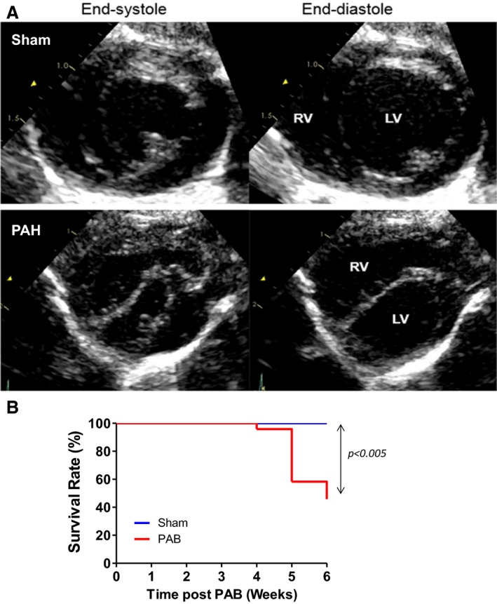
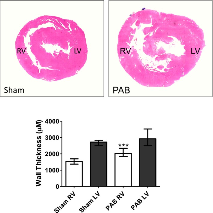
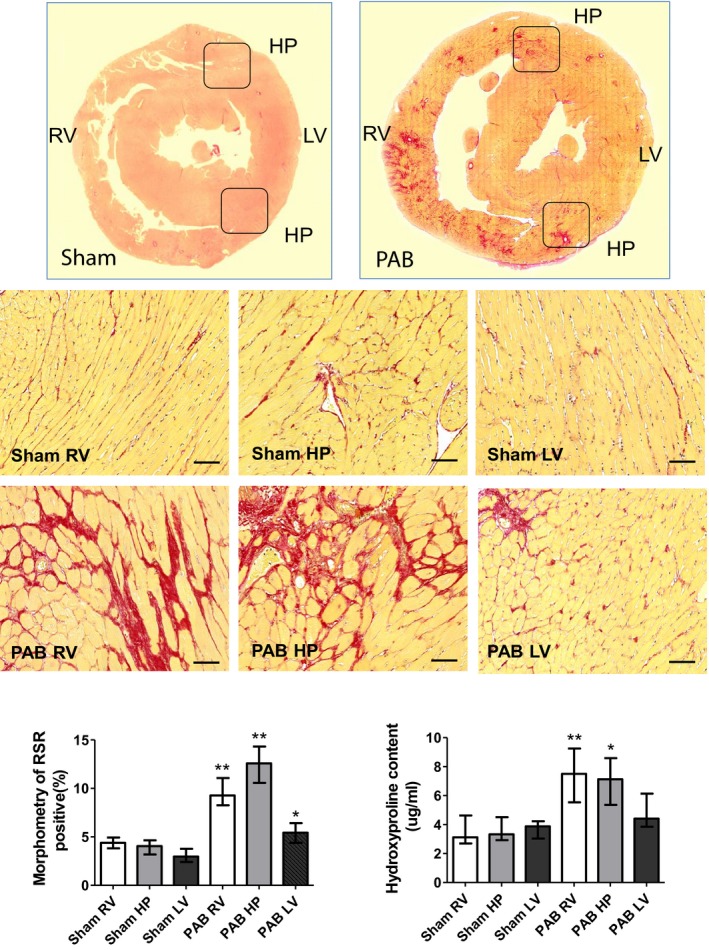
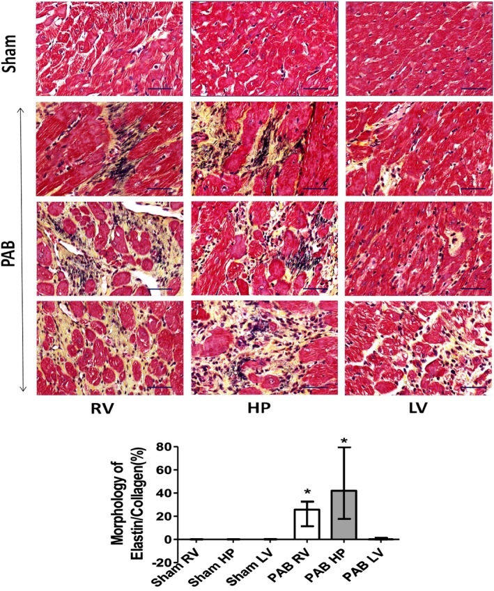
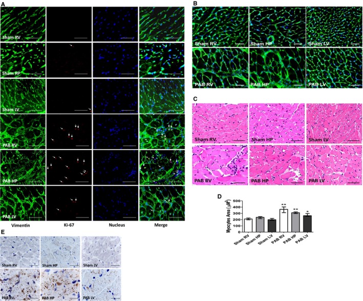
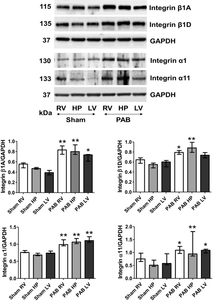
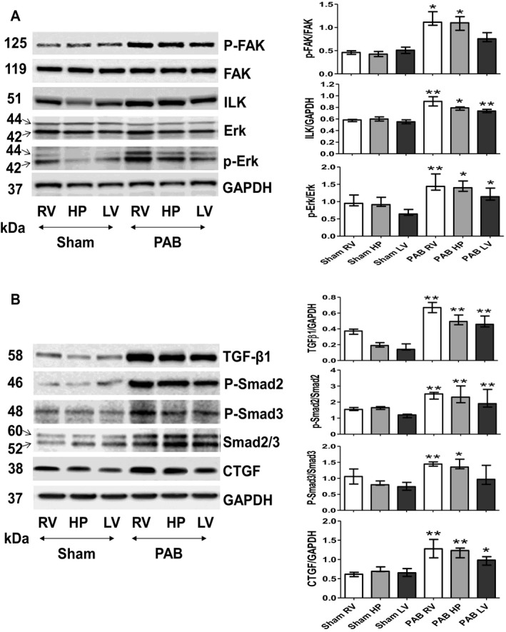
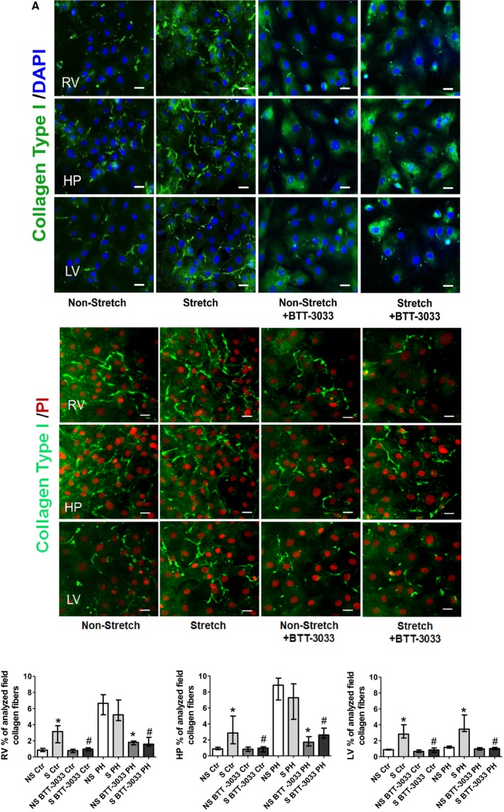
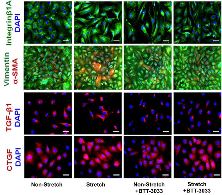
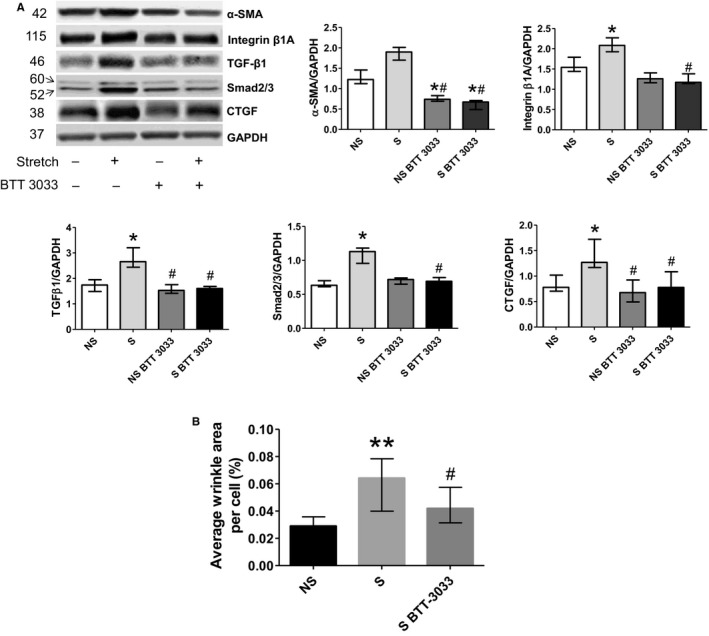
Methods and results: Pulmonary artery banding was used to induce RV hypertension in rats in vivo. Then, we evaluated cardiac function and regional remodeling 6 weeks after pulmonary artery banding. To further elucidate mechanisms responsible for regional cardiac remodeling, we also mimicked RV hypertensive stress by cyclic mechanical stretching applied to confluent cultures of cardiac fibroblasts, isolated from the RV free wall, septal hinge points, and LV free wall. Echocardiography and catheter evaluation demonstrated that rats in the pulmonary artery banding group developed RV hypertension with leftward septal displacement, LV compression, and increased LV end-diastolic pressures. Picrosirius red staining indicated that pulmonary artery banding induced marked RV fibrosis and dysfunction, with prominent fibrosis and elastin deposition at the septal hinge points but less LV fibrosis. These changes were associated with proportionally increased expressions of integrin-β1 and profibrotic signaling proteins, including phosphorylated Smad2/3 and transforming growth factor-β1. Moreover, mechanically stretched fibroblasts also expressed significantly increased levels of α-smooth muscle actin, integrin-β1, transforming growth factor-β1, collagen I deposition, and wrinkle formation on gel assays, consistent with myofibroblast transformation. These changes were not observed in parallel cultures of mechanically stretched fibroblasts, preincubated with the integrin inhibitor (BTT-3033).

Conclusions: Experimentally induced RV hypertension triggers regional RV, hinge-point, and LV integrin β1-dependent mechanotransduction signaling pathways that eventually trigger myocardial fibrosis via transforming growth factor-β1 signaling. Reduced LV fibrosis and preserved global function, despite geometrical and pressure aberrations, suggest a possible elastin-mediated protective mechanism at the septal hinge points.

Keywords: fibrosis; integrin; pressure overload; regional stress.


Figures

Figure 1
Figure 1
A, Representative echocardiography indicating that increasing the right ventricular (RV) pressure load induces RV dilatation, septal flattening, and left ventricular (LV) compression. Septal flattening changes geometry at the septal hinge‐point regions and LV. Short‐axis view obtained at the papillary muscle level at end systole and end diastole in a sham (A) and Pulmonary Arterial Hypertension (PAH) (B) rat. The sham rat shows a circular LV with normal round interventricular septal curvature and position throughout the cardiac cycle; in PAH, the RV is markedly enlarged and the LV is flattened and “D shaped” throughout the cardiac cycle, with the interventricular septum displaced leftward in systole and flattened into end diastole. B, Kaplan‐Meier survival curves in sham (n=8) and pulmonary artery banding (PAB) (n=26) at 6 weeks in rats. The median survival in PAB is 6. P<0.005 vs sham. Echocardiographic parameters are summarized in table 1.
Figure 2
Figure 2
Representative hematoxylin and eosin (H&E)–stained histologic morphometry of rat hearts 6 weeks after sham and pulmonary artery banding (PAB) procedures. H&E‐stained heart cross‐sections derived from sham and PAB animals (top). Morphometric quantification indicated that PAB induced a significantly increased right ventricular (RV) free‐wall thickness vs shams, whereas left ventricular (LV) free‐wall thickness was similar between groups. Values are expressed as medians and interquartile range (n=5). ***P<0.0001 vs sham.
Figure 3
Figure 3
Representative Picrosirius red (PSR) staining of rat hearts 6 weeks after sham and pulmonary artery banding (PAB) procedures. PSR staining demonstrates that PAB‐induced right ventricular (RV) pressure load is associated with a remarkable accumulation of PSR‐positive collagen and myocyte hypertrophy in the RV and septal hinge‐point (HP) regions. In contrast, the left ventricle (LV) displays only disseminated foci of PSR‐positive material (top panel). Low magnification of heart histological cross‐sections derived from sham and PAB animals and stained with PSR (middle panels). Higher magnification of representative sections derived from the RV, hinge‐point (HP), and LV heart regions (bottom panels). Bars=50 μm. The bar graphs depict morphometric quantification of the areas occupied by PSR‐positive collagen, as well as values of hydroxyproline content. Values are expressed as medians and interquartile range (n=5). *P<0.05, **P<0.005 vs sham.
Figure 4
Figure 4
Representative Movat's staining of rat hearts 6 weeks after sham and pulmonary artery banding (PAB) procedures. Movat's staining depicting collagen I (yellow) and elastin (black) marking differences in regional cardiac fibrosis and extracellular matrix composition in fragments dissected from the right ventricle (RV), hinge‐point (HP), and left ventricle (LV) heart regions of sham and PAB rats (top). There is increased elastin deposition most prominently at the septal HP. Bars=50 μm. The bar graphs depict morphometric quantification of areas occupied by Movat's‐positive elastin and collagen (bottom). Values are expressed as medians and interquartile range (n=5). *P<0.05 vs sham.
Figure 5
Figure 5
Representative micrographs of transverse sections of rat hearts sham and 6 weeks after pulmonary artery banding (PAB) procedures. A, Immunofluorescent detection of vimentin‐positive fibroblasts (green) and those displaying the presence of Ki‐67 proliferative antigen (red). Cell nuclei were stained blue, with 4′,6‐diamidino‐2‐phenylindole (DAPI). Bars=15 μm. B, Wheat germ agglutinin (WGA) interacting with cardiac myocyte cell membranes, detected with green fluorescein isothiocyanate (FITC; fluorescein) staining. Bar=20 μm. C, Hematoxylin and eosin staining for cardiomyocyte cross‐sectional area. Bar=20 μm. D, The bar graphs depict morphometric assessment of cardiac myocyte areas (n=5, with 200 cells per section). Values are expressed as medians and interquartile range. E, Immunohistochemical analysis of natriuretic peptide (NPPA) in cardiac muscle of sham and PAB rats. Bar=50 μm. HP indicates hinge point; LV, left ventricle; NPPA, Natural antisense transcript of natriuretic peptide precursor A; and RV, right ventricle. *P<0.05, **P<0.001 vs sham.
Figure 6
Figure 6
Right ventricular (RV) pressure load diversely upregulates levels of indicated integrins in heart regions 6 weeks after sham and pulmonary artery banding (PAB) procedures. Representative Western blots detecting indicated integrins and their quantitative assessments in the RV, hinge‐point (HP), and left ventricle (LV) regions of sham and PAB rats. Values are expressed as medians and interquartile range (n=5). *P<0.05, **P<0.001 vs sham.
Figure 7
Figure 7
Application of pulmonary artery banding (PAB) induces a diverse upregulation in activity of the integrin‐dependent signals that consequently trigger profibrotic pathway 6 weeks after sham and PAB procedures. A, Representative Western blots detecting basic regional expression of protein levels and phosphorylation rates of the integrin‐induced downstream signaling pathway components (focal adhesion kinase [FAK], integrin‐linked kinase [ILK], and extracellular signal–regulated kinase [ERK]). Values are expressed as medians and interquartile range (n=5). * P<0.05, ** P<0.001 vs sham. B, Representative Western blots detecting basic regional expression of protein levels and phosphorylation rates of profibrotic components transforming growth factor (TGF)‐β1, Smad 2, Smad 3, and connective tissue growth factor (CTGF) in sham and PAB‐exposed hearts. Quantification analyses (n=5). Values are expressed as medians and interquartile range. *P<0.05, **P<0.001 vs sham; . HP indicates hinge point; LV, left ventricle; and RV, right ventricle.
Figure 8
Figure 8
Cyclic mechanical stretch (24 hours long) of cultured cardiac fibroblasts isolated from the indicated heart regions. A, From control rats, induced a strong upregulation of immune‐detected collagen I deposition, compared with the nonstretched counterparts. This phenomenon has been suppressed in parallel cultures exposed to integrin inhibitor (BTT‐3033) (top panels). B, From pulmonary hypertension rats, indicated strong immune‐detected collagen I deposition in non‐stretched and stretched cells of right ventricular (RV) and hinge‐point (HP) regions, but not in left ventricle (LV). C, Representative fields of cardiac fibroblasts immune stained with collagen type I (green) and nuclear 4′,6‐diamidino‐2‐phenylindole (DAPI) staining (blue)/propidium iodide (PI) (red). Bar=50 μm. The bar graph represents the quantification data of the percentage of analyzed collagen fibers per field. Values are expressed as medians and interquartile range (n=4). Ctr indicates control; NS, nonstretch; PH, pulmonary hypertension; and S, stretch. * P<0.05 vs NS group; # P<0.05 vs S group.
Figure 9
Figure 9
Representative micrographs depicting cultures of cardiac fibroblasts isolated from right ventricle (RV) that were either kept still or subjected to 24‐hour‐long mechanical stretching in the presence or absence of integrin inhibitor (BTT‐3033). The parallel cultures were immune stained with specific antibodies recognizing integrin‐β1A (green), vimentin (green), α‐smooth muscle actin (α‐SMA) (red), transforming growth factor (TGF)‐β1 (red), and connective tissue growth factor (CTGF) (red), combined with blue 4′,6‐diamidino‐2‐phenylindole (DAPI) nuclear staining. Bars=50 μm.
Figure 10
Figure 10
Western blot assessment of profibrotic pathway components in right ventricular (RV) cardiac fibroblast cultures. A, Cyclic mechanical stretch of cultured cardiac fibroblasts causes upregulation in the net expressions of their α‐smooth muscle actin (α‐SMA), integrin‐β1, and profibrotic signaling pathway components (transforming growth factor [TGF]‐β1), Smad 2/3, and connective tissue growth factor [CTGF]). Western blots with indicated antibodies were assessed by densitometry and normalized against the GAPDH‐positive bands (n=4). Values are expressed as medians and interquartile range. *P<0.05 vs nonstretch (NS) group; # P<0.05 vs stretch (S) group. B, Percentage of analyzed collagen fibers and average cell surface wrinkles visible area to indicate cell contraction in gel assays. Values are expressed as medians and interquartile range (n=5). **P<0.005 vs NS group; # P<0.05 vs S group.

Similar articles

Cited by

References

    1. Badesch DB, Champion HC, Sanchez MA, Hoeper MM, Loyd JE, Manes A, McGoon M, Naeije R, Olschewski H, Oudiz RJ, Torbicki A. Diagnosis and assessment of pulmonary arterial hypertension. J Am Coll Cardiol. 2009;54:S55–S66. - PubMed
    1. Belenkie I, Horne SG, Dani R, Smith ER, Tyberg JV. Effects of aortic constriction during experimental acute right ventricular pressure loading: further insights into diastolic and systolic ventricular interaction. Circulation. 1995;92:546–554. - PubMed
    1. Mitchell JR, Whitelaw WA, Sas R, Smith ER, Tyberg JV, Belenkie I. RV filling modulates LV function by direct ventricular interaction during mechanical ventilation. Am J Physiol Heart Circ Physiol. 2005;289:H549–H557. - PubMed
    1. Damiano RJ Jr, La Follette P Jr, Cox JL, Lowe JE, Santamore WP. Significant left ventricular contribution to right ventricular systolic function. Am J Physiol. 1991;261:H1514–H1524. - PubMed
    1. Hirata M, Ousaka D, Arai S, Okuyama M, Tarui S, Kobayashi J, Kasahara S, Sano S. Novel model of pulmonary artery banding leading to right heart failure in rats. Biomed Res Int. 2015;2015:753210. - PMC - PubMed

Publication types

MeSH terms

LinkOut - more resources