. 2018 Mar 30;359(6383):1520-1523.
doi: 10.1126/science.aal2022.

Erythrocytic ferroportin reduces intracellular iron accumulation, hemolysis, and malaria risk

Affiliations

Erythrocytic ferroportin reduces intracellular iron accumulation, hemolysis, and malaria risk

De-Liang Zhang et al. Science. .

Abstract

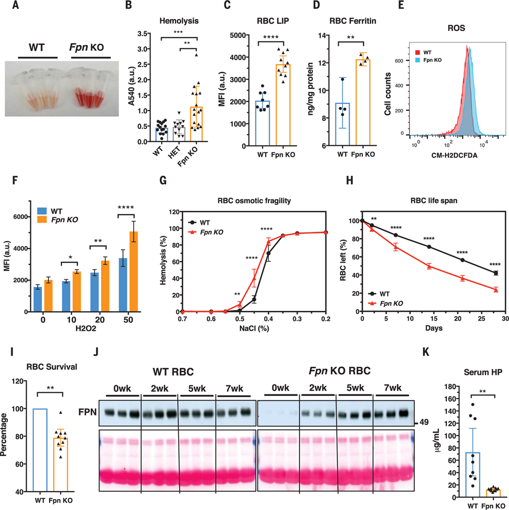
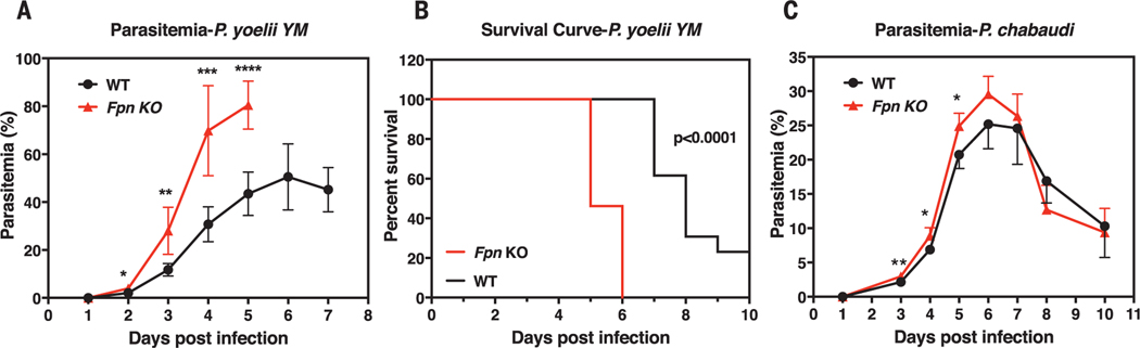
Malaria parasites invade red blood cells (RBCs), consume copious amounts of hemoglobin, and severely disrupt iron regulation in humans. Anemia often accompanies malaria disease; however, iron supplementation therapy inexplicably exacerbates malarial infections. Here we found that the iron exporter ferroportin (FPN) was highly abundant in RBCs, and iron supplementation suppressed its activity. Conditional deletion of the Fpn gene in erythroid cells resulted in accumulation of excess intracellular iron, cellular damage, hemolysis, and increased fatality in malaria-infected mice. In humans, a prevalent FPN mutation, Q248H (glutamine to histidine at position 248), prevented hepcidin-induced degradation of FPN and protected against severe malaria disease. FPN Q248H appears to have been positively selected in African populations in response to the impact of malaria disease. Thus, FPN protects RBCs against oxidative stress and malaria infection.


Figures

Fig. 1.
Fig. 1.. FPN is highly abundant on mature RBCs, and its activity is inhibited by iron supplementation and hepcidin.
(A) Immunoblots with four different FPN antibodies showed that FPN protein was present in membrane fractions of human and mouse RBCs. Actin is present as a loading control. (B) FPN protein levels were reduced in RBCs of erythroblast-specific Fpn KO mice. (C) FPN was about half as abundant in RBCs as in erythroblasts, but other iron metabolism proteins, including transferrin receptor 1 (TfR1), divalent metal transporter 1 (DMT1), and iron regulatory protein 1 (IRP1), were absent in RBCs (total lysate). Cells were purified by flow cytometry from bone marrow (fig. S2). Bottom image shows Ponceau S staining as a loading control. NonEryth, nonerythroblasts; Eryth, erythroblasts; Retic, reticulocytes. (D) Export of Fe55 by WT and Fpn KO RBCs was measured after 1 hour at 37°C, at 37°C with 1 μg/ml hepcidin (hep), or at 4°C. Mean ± 95% confidence interval (CI); n = 3. The experiments were independently repeated three times. Significance was determined by two-way analysis of variance (ANOVA) and Sidak’s multiple comparisons test. ****P < 0.0001; ns, not significant. (E) FPN levels were lower in membranes of RBCs from mice treated on high-iron diets (high) versus low-iron diets (low). Each lane represents a sample from one individual mouse. (F and G) FPN levels in total lysates of (F) erythroblasts and (G) RBCs treated ex vivo without (cont) or with 1 μg/ml hepcidin (hep) for 24 hours. Two independent replicates are shown in adjacent lanes. Numbers to the right of immunoblots are in kDa.
Fig. 2.
Fig. 2.. Fpn knockout in erythroblasts leads to RBC iron overload and intravascular hemolysis.
(A) Plasma of WT and Fpn KO mice after storing blood samples for 20 hours at 4°C showed increased hemolysis of Fpn KO RBCs. (B) Free-hemoglobin levels in plasma of WT, heterozygote (HET), and Fpn KO mice. Significance determined by one-way ANOVA and Tukey’s multiple comparisons test. A540, absorption at 540 nm; a.u., arbitrary units. (C) Labile iron pool (LIP) and (D) ferritin levels were dramatically increased in RBCs of Fpn KO mice. MFI, median fluorescence intensity. (E) Representative flow cytometry of reactive oxygen species (ROS) in RBCs of WT and Fpn KO mice and (F) quantification of ROS levels in RBCs of WT and Fpn KO mice after stimulation by H2O2 at different micromolar concentrations. n = 8. CM-H2DCFCA is a ROS indicator. (G) Osmotic fragility of RBCs, n = 9. (H) RBC life span of Fpn KO mice, n = 9. (I) Survival of ex vivo biotin-labeled WT and Fpn KO RBCs in the same WT mice (fig. S9). (J) Immunoblots showing FPN levels in the aging process of the RBCs of three WT and three Fpn KO mice in vivo. Sulfo-NHS-LC-Biotin was injected intravenously to label all RBCs, and after 0, 2, 5, and 7 weeks (wk), biotinylated RBCs were purified and FPN levels were then measured with immunoblots in total lysates. Ponceau S staining (bottom) is shown as a loading control. The increase of FPN levels in RBCs of Fpn conditional KO mice indicated that the few FPN-expressing RBCs survived longer than Fpn-null RBCs and were proportionally enriched over time (fig. S10). (K) Serum haptoglobin (HP) was depleted in Fpn KO mice. Data are presented as mean ± 95% CI. Significances for (F) to (H) were determined by two-way ANOVA and Sidak’s multiple comparisons test; significances for (I) and (K) were determined by Welch’s t test. *P < 0.05; **P < 0.01; ***P < 0.001; ****P < 0.0001.
Fig. 3.
Fig. 3.. FPN protects mice from severe malarial infection.
(A) Parasitemia (n = 5 for each group) and (B) survival curve (n = 13 for each group) of WT and Fpn KO mice after P. yoelii YM infection. (C) Parasitemia of WT and Fpn KO mice after P. chabaudi infection; n = 5 for WT and n = 7 for Fpn KO mice. Data are presented as mean ± 95% CI. Statistical significances determined for (A) and (C) with the Holm-Sidak multiple comparisons test with α = 0.05. Survival curves were analyzed with the log-rank test and Gehan-Breslow-Wilcoxon test. *P < 0.05; **P < 0.01; ***P < 0.001; ****P < 0.0001.

Similar articles

Cited by

References

    1. Cowman AF, Healer J, Marapana D, Marsh K, Cell 167, 610–624 (2016). - PubMed
    1. Sigala PA, Crowley JR, Hsieh S, Henderson JP, Goldberg DE, J. Biol. Chem 287, 37793–37807 (2012). - PMC - PubMed
    1. Clark MA, Goheen MM, Cerami C, Front. Pharmacol 5, 84 (2014). - PMC - PubMed
    1. Scholl PF, Tripathi AK, Sullivan DJ, Curr. Top. Microbiol. Immunol 295, 293–324 (2005). - PubMed
    1. Prentice AM, J. Nutr 138, 2537–2541 (2008). - PubMed

Publication types

MeSH terms