. 2018 Mar 30;359(6383):1527-1533.
doi: 10.1126/science.aar2094.

Molecular mechanism of extreme mechanostability in a pathogen adhesin

Affiliations

Molecular mechanism of extreme mechanostability in a pathogen adhesin

Lukas F Milles et al. Science. .

Abstract

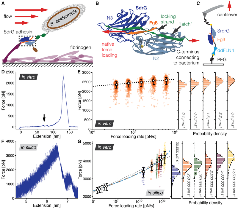
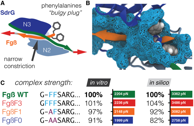
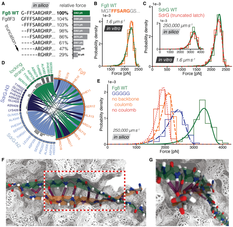
High resilience to mechanical stress is key when pathogens adhere to their target and initiate infection. Using atomic force microscopy-based single-molecule force spectroscopy, we explored the mechanical stability of the prototypical staphylococcal adhesin SdrG, which targets a short peptide from human fibrinogen β. Steered molecular dynamics simulations revealed, and single-molecule force spectroscopy experiments confirmed, the mechanism by which this complex withstands forces of over 2 nanonewtons, a regime previously associated with the strength of a covalent bond. The target peptide, confined in a screwlike manner in the binding pocket of SdrG, distributes forces mainly toward the peptide backbone through an intricate hydrogen bond network. Thus, these adhesins can attach to their target with exceptionally resilient mechanostability, virtually independent of peptide side chains.


Figures

Fig. 1
Fig. 1. The SdrG:Fgß complex withstands enormous forces in vitro and in silico.
(A) SdrG function, attached to the N-terminal peptide of the fibrinogen (purple) ß chain (orange) adsorbed on a surface. This interaction prevents detachment of the bacterium by hydrodynamic forces. (B)Structure of the SdrG (blue):Fgß (orange) complex. The locking strand (green) encloses the peptide in the binding pocket between the Ig-fold N2 (light blue) and N3 (dark blue) domain and a calcium (yellow) binding loop. The red arrows indicate the force applied to the molecular complex. (C) Experimental AFM setup including the ddFLN4 fingerprint domain (cyan). All constructs are covalently bound to the surface via Polyethyleneglycol (PEG) linkers and the ybbR-tag (yellow dots). In the native configuration, Fgß and SdrG are force loaded from their respective C-termini. The AFM cantilever is retracted at constant velocity until the complex breaks. (D) Resulting force-extension trace in the native force propagation (blue), as it would occur at sites of staphylococcal adhesion. The distinctive fingerprint unfolding around 90 pN ddFLN4 (black arrow) featuring a substep was used to find specific interactions. It is followed by SdrG:Fgß complex rupture, here at almost 2500 pN. (E) Dynamic force spectrum of the SdrG:Fgß native geometry at cantilever retraction velocities 0.4 μm s−1 (triangles, N = 749), 0.8 μm s−1 (squares, N = 696), 1.6 μm s−1 (diamonds, N = 758), 3.2 μm s−1 (forward triangles, N = 749), 6.4 μm s−1 (circles, N = 851), with corresponding complex rupture force histograms for each velocity projected onto individual axes on the right. A Bell-Evans (BE) model fit (dotted line, Δx = 0.051 nm, koff0 = 9.2E-11s−1) through the most probable rupture force and force loading rate of each velocity (large open markers, with errors given as full-width at half maximum for each distribution) shows the expected force loading-rate dependency of the rupture force. (F) SMD force-extension trace (blue) in the native force propagation of SdrG:Fgß including experimental peptide linkers. The complex ruptured at almost 4000 pN, the extension is shorter than in the experimental trace, as there are no PEG spacers. The peak following the highest force peak corresponds to another metastable geometry after slipping of the Fgß peptide, that is below the resolution limit of our AFM. (G) The experimentally determined dynamic force spectrum from velocities of 0.4 to 6.4 μm s−1 for the native propagation from (E) is shown condensed as open circles. The dynamic force spectrum of steered MD simulations for velocities of (25,000 μm s−1 to 12,500,000 μm s−1, triangle N = 49, square N = 50, diamond N = 100, forward triangle N = 200, pentagon N = 147, inverted triangle N = 200, respectively). Fits through SMD and experimental data, for BE model (gray, dotted line, Δx = 0.047 nm, koff0 = 1.0E-9 s−1) and fit of a model by Dudko et. al. (DHS model, cusp potential Δx = 0.12 nm, koff0 = 6.1E-22 s−1, ΔG++ = 78 kBT, cyan dashed line and linear-cubic potential Δx = 0.093 nm, koff0 = 7.7E-18 s−1, ΔG++ = 66 kBT, brown dash-dotted line, both at T = 300 K). In vitro and in silico data agree exceptionally well, although they are separated by six orders of magnitude in force loading rate and can be fit with a single model.
Fig. 2
Fig. 2. Phenylalanine side chains only marginally influence SdrG:Fgß mechanostability.
(A) Sketch of the “bulgy plug” hypothesis. The bulky phenyalanine side chains (gray) of Fgß (orange) are blocked by the locking strand (green). (B) Crystal structure showing the bulky phenylalanine sidechains in van der Waals representation (gray spheres) of Fgß (orange). They have to wiggle through a narrow constriction (cyan surface). (C) Dependence of complex rupture force on the presence of phenylalanines, if replaced by alanines. Most probable rupture forces (absolute values in bar graphs) are compared relative to WT Fgß. Either recorded experimentally with a single cantilever retracted at 1.6 μm s−1 or corresponding results for SMD simulations at 250,000 μm s−1. Adding one F (FgßF3 mutant) slightly increases forces. Yet, both results show a trend of weak dependence of rupture force on the presence of phenylalanines. Even when removing all bulky side chains (FgßF0 mutant) experimental rupture forces drop no more than 10% compared to WT Fgß, in silico no more than 20%. The “bulgy plug” only marginally contributes, hinting that another mechanism must be responsible for the high forces.
Fig. 3
Fig. 3. Backbone H-bonds are deciding factors in the high mechanostability of Sdrg:Fgß and a minimized peptide.
(A) Fgß peptide truncations from the N-terminus in silico. By removing amino acids the forces drop (relative to the WT) with the most significant drop when removing the sequence FSAR, leading to FFSARG as the minimum peptide. (B) Rupture forces for SdrG binding to WT Fgß (green, continuous line, N = 437), and the six-residue minimized peptide FFSARG (orange, dash-dotted line, here shown with surrounding amino acids in gray, N = 471). Strikingly, there is hardly any difference between WT Fgß and the minimized peptide. (C) Rupture force histograms comparing the wildtype Fgß:SdrG interaction (green, continuous line, N = 463), and the SdrG mutant with the truncated latch region (red, dashed line, N = 131). WT and mutant are virtually indistinguishable (no significant difference in Kolmogorov–Smirnov test, in vitro p = 0.29, in silico p = 0.88). Corresponding SMD results WT N = 100, mutant N = 50) are shown as inset. (D) Relative prevalence (bar graphs, precise values in Fig. S7) of H-bonds between SdrG domains, the locking strand and the WT Fgß peptide (also available for F3, F1, F0, and all-Glycine mutants in Fig. S8). The locking strand connects to nearly every Fgß residue. (E) Rupture forces from exploratory simulations for SdrG and Fgß WT (green, continuous line, N = 100), a replacement of each Fgß residue with glycine (blue, dash-dotted line, N = 100), FgßF3 peptide without coulomb interactions, and subsequently H-bonds, on its backbone (orange, dashed line, N = 47), FgßF3 devoid of all coulomb interactions (red, dotted line, N = 48). Backbone H-bonds in the Fgß confinement allow even a pure glycine sequence to withstand high force. (F) H-bond (purple) contacts respective to the backbone of Fgß (orange) and locking strand (green) confined by SdrG (white surface) from simulations in a force loaded state. The minimum peptide sequence is highlighted in the red box (G) Radial distribution of backbone H-bonds between locking strand (green) caused by the screw-like winding of the Fgß sheet (orange). Peptide backbones are shown as sticks.
Fig. 4
Fig. 4. A non-native SdrG:Fgß force loading shows weak forces, a homologous domain ClfB reaches 2 nN stability binding a mainly glycine-serine peptide and SdrG homologs consistenly exceed 2 nN binding to their ligands.
(A) Dynamic force spectrum of the SdrG:Fgß non-native configuration, see inset with purple arrow, breaking around 60 pN as opposed to > 2 nN for the native case (for SMD results see Fig. 15-17 and video S22). Cantilever retraction velocities were varied: 0.4 μm s−1 (triangles, N = 511), 0.8 μm s−1 (squares, N = 564), 1.6 μm s−1 (diamonds, N = 487), 3.2 μm s−1 (forward triangles, N = 395), 6.4 μm s−1 (circles, N = 471) with corresponding complex rupture force histograms projected on the right. A BE model fit (dashed line) through the most probable rupture force and force loading rate of each velocity (large open markers) shows the expected force loading-rate dependency of the rupture force Δx = 0.46 nm, koff0 = 0.39 s−1). (B) ClfB (blues):K10 (orange) complex including the locking strand (green) and H-bonding (purple) amino acids shown as sticks. Notably, the latch region was not crystallized and needed to be modeled from a homolog. The native pulling configuration is indicated with an arrow, compared to Fgμ the peptide is oriented inversely in the binding pocket. (C) Rupture force histogram and fit for ClfB:K10 at a velocity of 0.8 μm s−1 (green, dashed line, N = 1035) peaking around 2.3 nN. Simulation data (N = 50) confirming the force regime are shown as inset. (D) Homologous systems employing the DLL mechanism, all from S. aureus (N2 and N3 domains in blue, target peptides in orange) SdrE, Bbp, FnBPA, and ClfA (E) Comparison of absolute mechanostability of all homologous systems as well as SdrG and ClfB with a single force probe. The cantilever is modified with five different peptides tethered in their native force loading geometry, respectively: from the C-terminus of Complement Factor H (CFH), Fgα chain (Fgα), and Fgß, tethered from the N-terminus are sequences from Dermokine (DK), and Fgγ chain (Fgγ). This selection is presented to all adhesins, which are known to bind at least one of them, spatially separated on a surface. One cannot exclude that one adhesin may bind more than one peptide target. (F) Resulting relative stabilities of the complexes for SdrE (red, dashed line, N = 680), ClfB (orange, dash-dotted line, N= 605), ClfA (cyan, dashed line, N = 2292), Bbp (purple, dot-dot-dashed line, N = 319), SdrG (green, continuous line, N = 478), FnBPA (blue, dash-dash-dotted line, N = 2483). SdrG is not the strongest system at a retraction velocity of 1.6 μm s−1. In accordance with the largely side-chain independent mechanics proposed for SdrG and ClfB, every DLL adhesin withstands forces exceeding 2 nN.

Comment in

Similar articles

Cited by

References

    1. Foster TJ, Geoghegan JA, Ganesh VK, Höök M, Adhesion, invasion and evasion: the many functions of the surface proteins of Staphylococcus aureus. Nat. Rev. Microbiol 12, 49–62 (2013). - PMC - PubMed
    1. Otto M, Staphylococcus epidermidis — the “accidental” pathogen. Nat. Rev. Microbiol 7, 555–567 (2009). - PMC - PubMed
    1. Deivanayagam CCS et al., A novel variant of the immunoglobulin fold in surface adhesins of Staphylococcus aureus: Crystal structure of the fibrinogen-binding MSCRAMM, clumping factor A. EMBO J. 21, 6660–6672 (2002). - PMC - PubMed
    1. Ponnuraj K et al., A “dock, lock, and latch” Structural Model for a Staphylococcal Adhesin Binding to Fibrinogen. Cell. 115, 217–228 (2003). - PubMed
    1. Ganesh VK et al., Structural and Biochemical Characterization of Staphylococcus aureus Clumping Factor B/Ligand Interactions. J. Biol. Chem 286, 25963–25972 (2011). - PMC - PubMed

Publication types