Review
. 2018 Jul 6;293(27):10524-10535.
doi: 10.1074/jbc.TM118.000372. Epub 2018 Mar 29.

Homologous recombination and the repair of DNA double-strand breaks

Affiliations
Review

Homologous recombination and the repair of DNA double-strand breaks

William Douglass Wright et al. J Biol Chem. .

Abstract

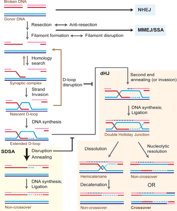
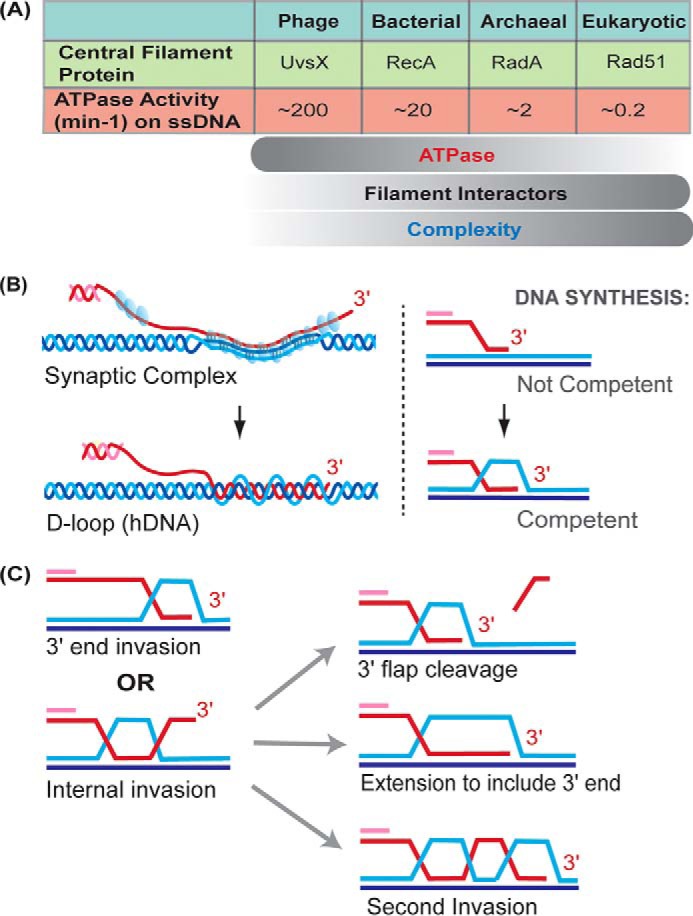
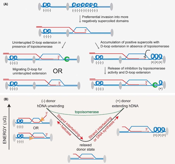
Homologous recombination enables the cell to access and copy intact DNA sequence information in trans, particularly to repair DNA damage affecting both strands of the double helix. Here, we discuss the DNA transactions and enzymatic activities required for this elegantly orchestrated process in the context of the repair of DNA double-strand breaks in somatic cells. This includes homology search, DNA strand invasion, repair DNA synthesis, and restoration of intact chromosomes. Aspects of DNA topology affecting individual steps are highlighted. Overall, recombination is a dynamic pathway with multiple metastable and reversible intermediates designed to achieve DNA repair with high fidelity.

Keywords: DNA damage; DNA endonuclease; DNA helicase; DNA polymerase; DNA recombination; DNA repair; DNA topoisomerase; DNA topology; genomic instability.


Conflict of interest statement

The authors declare that they have no conflicts of interest with the contents of this article

Figures

Figure 1.
Figure 1.
Model for repair of DNA double-strand breaks by homologous recombination in somatic cells. When a DNA double-strand break (DSB) occurs in a DNA molecule, repair can proceed by multiple pathways largely controlled by end resection. NHEJ is capable of repairing unresected or minimally resected DSBs in a template-independent fashion. MMEJ and single-strand annealing (SSA) rely on different extents of homology between the two DSB ends for repair independent of a donor molecule. Homologous recombination proceeds as shown in the figure using a homologous donor DNA. Most of the extended D-loops in somatic cells are disrupted and subsequently repaired by SDSA. The end result of the repair by SDSA is always a noncrossover outcome, thus avoiding loss of heterozygosity produced by somatic crossovers. SDSA occurs by disruption of the extended D-loop and annealing the newly synthesized DNA with the second end of the broken molecule. Alternatively, the newly synthesized strand may invade the second end as depicted in Fig. S1. The extended D-loop can also undergo second-end capture or invasion (Fig. S2) to form a double Holliday junction (dHJ). This may either lead to a crossover or a noncrossover outcome. Invasion by the second break end makes dHJ formation and hence crossover outcome more likely, as depicted in Fig. S2. See Fig. S3 for another model for crossover generation. dHJs can be dissolved into noncrossovers by the concerted action of the Sgs1–Top3–Rmi1 complex to migrate the two junctions toward each other and then decatenate the strands of the hemicatenane by the Top3 topoisomerase activity. Each colored line indicates a strand of DNA, and dotted lines represent DNA synthesis.
Figure 2.
Figure 2.
Homology search and DNA strand invasion. A, ATPase activity of central filament proteins decreases with organism complexity while filament interactors increase in number. From T4 phage UvsX to Rad51, the ssDNA-dependent ATPase activity decreases roughly four orders of magnitude. B, hypothetical cartoon of the synaptic complex intermediate. The arrangement of DNA strands within the Rad51 filament (blue spheres) is not known, but there is no net intertwining of DNA strands. Bases are shown paired in triplets based on the RecA crystal structure (33). Watson-Crick base pairing occurs between the invading strand and its complement and may be partially retained between the two complementary strands of the donor molecule. Intertwining of DNA strands leads to D-loop formation, containing heteroduplex DNA (hDNA). C, two types of invasion are 3′ end invasion, which is favored (81), and internal invasion away from the end. In the latter case, the D-loop must be processed to create a primer–template junction containing the 3′ end of the invading DNA strand for extension by a DNA polymerase, and different possibilities are depicted.
Figure 3.
Figure 3.
Topological considerations in DNA strand invasion. A, supercoiling density in various chromatin domains may influence the propensity to invade (invasion is a term inclusive of synaptic complex formation and hDNA formation through interwining of the invading strand) a particular domain in the donor. Synaptic complex formation initially relaxes the donor when the extended Rad51–ssDNA filament aligns with homologous bases present in the complementary donor strand, causing them to extend as well. Invading strand intertwining to produce hDNA is a separate consumption of the negative supercoils in the donor, and unlike synaptic complexes, hDNA is stable even in the absence of the bound proteins. The topological status of the donor will also influence the DNA synthesis step, as indicated in the figure and discussed in the text. Gray ovals indicate a specific DNA-bound protein(s), which creates discrete topological domains within linear chromosomes. Green horseshoe, topoisomerase. B, negative supercoiling creates an energy well that influences DNA strand intertwining or unwinding. Invasion into a supercoiled donor will favor stand intertwining as negative supercoils release their stored energy as they are dissipated, with a relaxed donor defining the lowest energy state. Once all negative supercoils are relaxed, extending hDNA through further intertwining, or DNA synthesis, will induce positive supercoils. Unwinding the D-loop in a relaxed donor by a DNA helicase will require they rewind negative supercoils in the donor. The dotted green line represents the altered energy landscape made possible by the action of topoisomerases, in effect making the energy well more shallow. Orange triangles depict DNA helicases and their translocation orientation to disrupt D-loops.

Similar articles

Cited by

References

    1. Kowalczykowski S. C., Hunter N., and Heyer W.-D. (eds) (2016) DNA Recombination, Cold Spring Harbor Laboratory Press, Cold Spring Harbor, NY
    1. Hustedt N., and Durocher D. (2016) The control of DNA repair by the cell cycle. Nat. Cell Biol. 19, 1–9 10.1038/ncb3452 - DOI - PubMed
    1. Pannunzio N. R., Watanabe G., and Lieber M. R. (2018) Nonhomologous DNA end-joining for repair of DNA double-strand breaks. J. Biol. Chem. 293, 10512–10523 10.1074/jbc.TM117.000374 - DOI - PMC - PubMed
    1. Sallmyr A., and Tomkinson A. (2018) Repair of DNA double-strand breaks by mammalian alternative end-joining pathways. J. Biol. Chem. 293, 10536–10546 10.1074/jbc.TM117.000375 - DOI - PMC - PubMed
    1. Her J., and Bunting S. F. (2018) How cells ensure correct repair of DNA double-strand breaks. J. Biol. Chem. 293, 10502–10511 10.1074/jbc.TM118.000371 - DOI - PMC - PubMed

Publication types