. 2018 May 25;293(21):8242-8254.
doi: 10.1074/jbc.RA117.001299. Epub 2018 Mar 29.

TGFβ and IGF1R signaling activates protein kinase A through differential regulation of ezrin phosphorylation in colon cancer cells

Affiliations

TGFβ and IGF1R signaling activates protein kinase A through differential regulation of ezrin phosphorylation in colon cancer cells

Premila D Leiphrakpam et al. J Biol Chem. .

Abstract

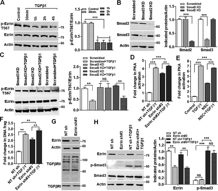
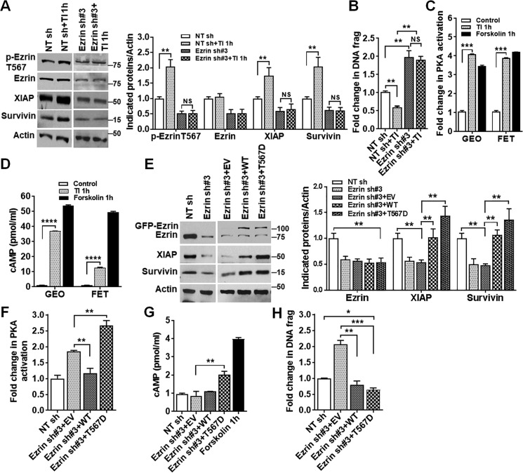
Aberrant cell survival plays a critical role in cancer progression and metastasis. We have previously shown that ezrin, a cAMP-dependent protein kinase A-anchoring protein (AKAP), is up-regulated in colorectal cancer (CRC) liver metastasis. Phosphorylation of ezrin at Thr-567 activates ezrin and plays an important role in CRC cell survival associated with XIAP and survivin up-regulation. In this study, we demonstrate that in FET and GEO colon cancer cells, knockdown of ezrin expression or inhibition of ezrin phosphorylation at Thr-567 increases apoptosis through protein kinase A (PKA) activation in a cAMP-independent manner. Transforming growth factor (TGF) β signaling inhibits ezrin phosphorylation in a Smad3-dependent and Smad2-independent manner and regulates pro-apoptotic function through ezrin-mediated PKA activation. On the other hand, ezrin phosphorylation at Thr-567 by insulin-like growth factor 1 receptor (IGF1R) signaling leads to cAMP-dependent PKA activation and enhances cell survival. Further studies indicate that phosphorylated ezrin forms a complex with PKA RII, and dephosphorylated ezrin dissociates from the complex and facilitates the association of PKA RII with AKAP149, both of which activate PKA yet lead to either cell survival or apoptosis. Thus, our studies reveal a novel mechanism of differential PKA activation mediated by TGFβ and IGF1R signaling through regulation of ezrin phosphorylation in CRC, resulting in different cell fates. This is of significance because TGFβ and IGF1R signaling pathways are well-characterized tumor suppressor and oncogenic pathways, respectively, with important roles in CRC tumorigenesis and metastasis. Our studies indicate that they cross-talk and antagonize each other's function through regulation of ezrin activation. Therefore, ezrin may be a potential therapeutic target in CRC.

Keywords: A-kinase anchoring protein (AKAP); IGF1R; Smad3; X-linked inhibitor of apoptosis protein (XIAP); apoptosis; colon cancer; ezrin; metastasis; survivin; transforming growth factor beta (TGF-β).


Conflict of interest statement

The authors declare that they have no conflicts of interest with the contents of this article

Figures

Figure 1.
Figure 1.
Knockdown of ezrin expression activates PKA and induces apoptosis in colon cancer cells. A, GEO and FET colon cancer cells with stable expression of ezrin shRNA (sh#1 and sh#3) showed a significant reduction in ezrin protein expression. Nontargeting shRNA (NT sh) was used as a control (left panel). Quantification of Western blots was performed as described under “Experimental procedures” (right panel). **, p < 0.01 (n = 3). B, ezrin KD cells showed an ∼2-fold increase in apoptosis, as determined by DNA fragmentation assays. **, p < 0.01 (n = 3). C, ezrin KD led to down-regulation of XIAP and survivin and an increase in cleaved caspase 7 (left panel). Quantification of Western blots is shown on the right. **, p < 0.01; ***, p < 0.001 (n = 4). D, ezrin KD GEO and FET cells exhibited increased PKA activation compared with NT sh cells. Forskolin (10 μm) was used as a positive control. **, p < 0.01 ; ***, p < 0.001 (n = 3). E, ezrin KD-mediated increase of p-CREB is abrogated by PKA inhibitor H89 (15 μm) (left panel). Quantification of Western blots is shown on the right. **, p < 0.01; ***, p < 0.001. NS, not statistically significant (n = 2). F, IP assays showed the decreased association of PKARII with PKACatα in ezrin KD cells compared with NT sh control cells (left panel). Relative levels of PKACatα to PKARII were analyzed, and the results are shown on the right. ***, p < 0.001 (n = 2). G, production of cAMP was not increased in ezrin KD cells as determined by cAMP assay. Forskolin was used as a positive control. NS, not statistically significant (n = 3); IB, immunoblot.
Figure 2.
Figure 2.
Knockdown of ezrin expression induces apoptosis through PKA activation. A, expression of PKA catalytic α subunit (PKACatα) was knocked down in FET cells, with no change in ezrin phosphorylation at Thr-567 and XIAP and survivin expression (left panel). Quantification of Western blots is shown on the right. **, p < 0.01. NS, not statistically significant (n = 3). B, PKACatα KD blocked endogenous as well as ezrin KD-mediated PKA activation. **, p < 0.01; ***, p < 0.001. NS, not statistically significant (n = 3). C, PKACatα KD abrogates ezrin KD-mediated XIAP and survivin down-regulation (left panel). Quantification of Western blots is shown on the right. **, p < 0.01. NS, not statistically significant (n = 3). D, PKACatα KD abrogates ezrin KD-induced apoptosis. ***, p < 0.001. NS, not statistically significant (n = 3).
Figure 3.
Figure 3.
Dephosphorylation of ezrin at Thr-567 regulates PKA activation and apoptosis in GEO cells. A, ezrin T567A down-regulated XIAP and survivin expression and increased caspase 7 cleavage (left panel). Quantification of Western blots is shown on the right. **, p < 0.01 (n = 3). B, ezrin T567A increased apoptosis. **, p < 0.01 (n = 3). C and D, ezrin T567A activated PKA with no increase in cAMP production. Forskolin was used as a positive control. **, p < 0.01. NS, not statistically significant (n = 3).
Figure 4.
Figure 4.
Inhibition of ezrin phosphorylation at Thr-567 by NSC668394 induces apoptosis through PKA activation. A and B, GEO and FET colon cancer cells treated with the ezrin inhibitor NSC668394 (NSC) showed a dose-dependent decrease in phosphorylation of ezrin at Thr-567, down-regulation of XIAP, and survivin expression (A, upper panel) and induction of apoptosis (B). The lower panels show quantification of Western blots. **, p < 0.01, ***, p < 0.001 (n = 3). C, PKA activity assays showed PKA activation by NSC (20 μm) treatment. PKA inhibitor H89 (15 μm) pretreatment abrogated endogenous and NSC-induced PKA activation. Forskolin was used as a positive control. ***, p < 0.001. NS, not statistically significant (n = 3). D, NSC-induced PKA activation is independent of cAMP production. NS, not statistically significant (n = 3). E, PKACatα KD blocked NSC-mediated XIAP and survivin down-regulation (upper panel). The lower panel shows quantification of Western blots. **, p < 0.01. NS, not statistically significant (n = 3). F and G, PKACatα KD abrogated NSC-mediated apoptosis (F) and PKA activation (G). **, p < 0.01. NS, not statistically significant (n = 3).
Figure 5.
Figure 5.
TGFβ inhibits ezrin phosphorylation at Thr-567, resulting in PKA activation and inhibition of XIAP and survivin expression. A, TGFβ1 treatment of FET cells decreased ezrin phosphorylation at Thr-567 in a time-dependent manner (left panel). Relative levels of p-ezrin Thr-567 to total ezrin were analyzed, and the results are shown on the right. ***, p < 0.001 (n = 3). B, expression of Smad2 and Smad3 was knocked down in FET cells (left panel). Quantification of Western blots is shown on the right. ***, p < 0.001 (n = 2). C, Smad3 KD, but not Smad2 KD, prevented TGFβ1-mediated inhibition of ezrin phosphorylation (left panel). Relative levels of p-ezrin Thr-567 to total ezrin were determined and are shown on the right. **, p < 0.01. NS, not statistically significant (n = 3). D, TGFβ1 treatment increased PKA activation to a much lesser degree in ezrin KD cells than in NT sh control cells. **, p < 0.01; ***, p < 0.001 (n = 3). E, NSC attenuated TGFβ-induced PKA activation. ***, p < 0.001 (n = 3). F, ezrin KD reduces TGFβ1-induced apoptosis. **, p < 0.01 (n = 2). G and H, ezrin KD has no effect on TGFβRI or TGFβRII expression (G) or on Smad3 expression or TGFβ-mediated Smad3 phosphorylation (H, left). Quantification of Western blots is shown on the right. **, p < 0.01; ***, p < 0.001. NS, not statistically significant (n = 3).
Figure 6.
Figure 6.
Hyperphosphorylation of ezrin at Thr-567 activates PKA and promotes survival. A, TI treatment increased phosphorylation of ezrin at Thr-567 and expression of XIAP and survivin together with inhibition of apoptosis in NT sh control cells. However, the increase was abrogated in ezrin KD cells (left panel). Quantification of Western blots is shown on the right. **, p < 0.01. NS, not statistically significant (n = 3). B, TI treatment reduced apoptosis in NT sh control cells, and this inhibition was abrogated in ezrin KD cells. **, p < 0.01. NS, not statistically significant (n = 2). C and D, TI activated PKA (C) and increased cAMP production (D) ***, p < 0.001; ****, p < 0.0001 (n = 3). E, ectopic expression of ezrin Thr-567 phospho-mimetic mutant (T567D) or WT ezrin increased XIAP and survivin expression in GEO cells (left panel). Quantification of Western blots is shown on the right. **, p < 0.01 (n = 3). F and G, ezrin T567D mutant enhanced PKA activation (F) concomitant with increased cAMP production (G). Forskolin is used as a positive control. **, p < 0.01 (n = 3). H, ectopic expression of ezrin T567D and ezrin WT reduced apoptosis. *, p < 0.05 and **, p < 0.01; ***, p < 0.001 (n = 3).
Figure 7.
Figure 7.
PKA activation induced by ezrin inhibition depends on AKAP149 expression. A, pretreatment with a pan AKAP inhibitor Ht31 (25 μm) abrogated NSC-mediated PKA activation in FET and GEO cells. **, p < 0.01; ***, p < 0.001. NS, not statistically significant (n = 3). B, AKAP149 KD had no effect on expression of ezrin, p-ezrin Thr-567, XIAP, or survivin but restored XIAP and survivin expression by ezrin KD (upper left panels). Ezrin KD had no effect on AKAP149 expression (upper right panel). Quantification of Western blots is shown in the lower panel. **, p < 0.01. NS, not statistically significant (n = 3). C, AKAP149 KD abrogated ezrin KD-mediated apoptosis. **, p < 0.01. NS, not statistically significant (n = 3). D, AKAP149 KD blocked NSC-mediated PKA activation. **, p < 0.01. NS, not statistically significant (n = 3). E, reciprocal IP analysis with AKAP149 or PKARII antibodies showed increased association of AKAP149 with PKARII in ezrin T567A cells and decreased association in ezrin T567D cells (upper panel). Relative levels of PKARII and AKAP149 were analyzed, and the results are shown in the lower panel. *, p < 0.05; **, p < 0.01 (n = 2). F, IP analysis with ezrin or PKARII antibodies indicated that there was more PKARII associated with ezrin in T567D cells than in T567A cells (upper panel). The relative levels of PKARII and ezrin are shown in the lower panel. *, p < 0.05; **, p < 0.01 (n = 2).
Figure 8.
Figure 8.
Proposed model of cross-talk between TGFβ–Smad3 and IGF1R signaling pathways. TGFβ–Smad3 inhibits ezrin phosphorylation at Thr-567, leading to the association of PKA RII with AKAP149 to activate PKA in a cAMP-independent manner, which suppresses XIAP and survivin expression and induces apoptosis. With IGF1R activation, ezrin is hyperphosphorylated at Thr-567, resulting in association of ezrin with PKA RII and PKA activation in a cAMP-dependent manner leading to XIAP and survivin up-regulation and cell survival. GF, growth factors; P, phosphorylation.

Similar articles

Cited by

References

    1. Markowitz S. D., and Bertagnolli M. M. (2009) Molecular origins of cancer: Molecular basis of colorectal cancer. N. Engl. J. Med. 361, 2449–2460 10.1056/NEJMra0804588 - DOI - PMC - PubMed
    1. André T., Boni C., Mounedji-Boudiaf L., Navarro M., Tabernero J., Hickish T., Topham C., Zaninelli M., Clingan P., Bridgewater J., Tabah-Fisch I., de Gramont A., and Multicenter International Study of Oxaliplatin/5-Fluorouracil/Leucovorin in the Adjuvant Treatment of Colon Cancer (MOSAIC) Investigators (2004) Oxaliplatin, fluorouracil, and leucovorin as adjuvant treatment for colon cancer. N. Engl. J. Med. 350, 2343–2351 10.1056/NEJMoa032709 - DOI - PubMed
    1. Mehlen P., and Puisieux A. (2006) Metastasis: a question of life or death. Nat. Rev. Cancer 6, 449–458 10.1038/nrc1886 - DOI - PubMed
    1. Nguyen D. X., Bos P. D., and Massagué J. (2009) Metastasis: from dissemination to organ-specific colonization. Nat. Rev. Cancer 9, 274–284 10.1038/nrc2622 - DOI - PubMed
    1. Leiphrakpam P. D., Rajput A., Mathiesen M., Agarwal E., Lazenby A. J., Are C., Brattain M. G., and Chowdhury S. (2014) Ezrin expression and cell survival regulation in colorectal cancer. Cell Signal. 26, 868–879 10.1016/j.cellsig.2014.01.014 - DOI - PMC - PubMed

Publication types

MeSH terms

LinkOut - more resources