

Large intra-abdominal seminoma in a left undescended testicle complicated by torsion
- PMID: 29599382
- PMCID: PMC5878376
- DOI: 10.1136/bcr-2017-222670
Large intra-abdominal seminoma in a left undescended testicle complicated by torsion
Abstract
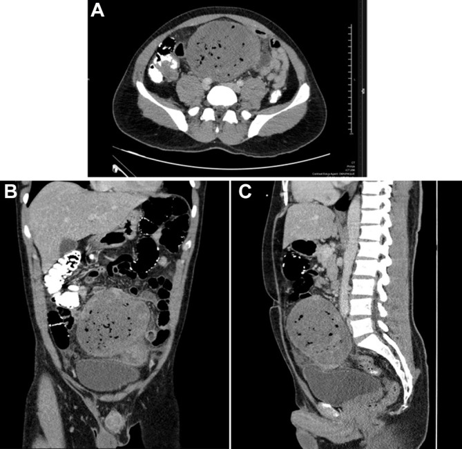
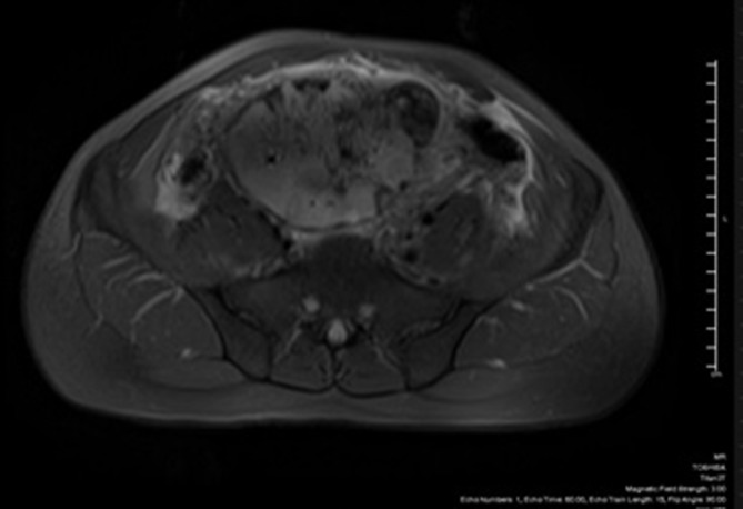
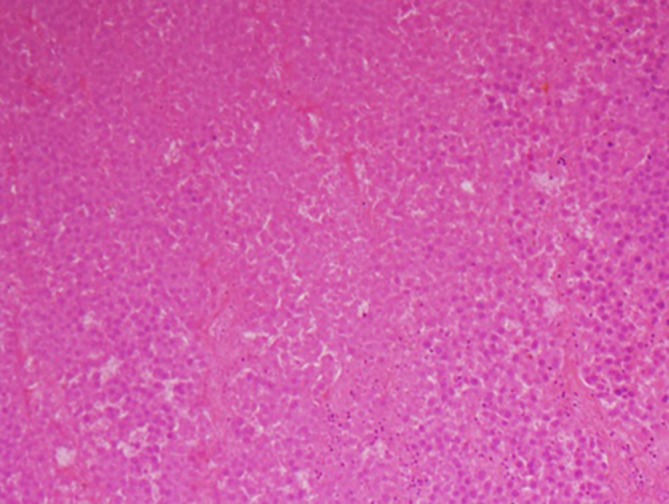
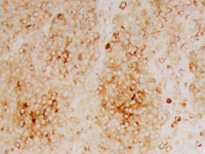
A 39-year-old man presented with a 2-day history of worsening constant, dull diffuse lower abdominal pain with associated constipation and known history of left undescended testicle. He was evaluated at an outside hospital where a non-contrasted CT scan revealed a 20 cm well-circumscribed soft tissue mass within the pelvis.He was referred and further imaging revealed a 12 cm heterogeneous mass with foci of air that appeared to be contiguous with the left spermatic cord. This constellation of findings could represent torsion of undescended left testicle with infarction or underlying malignancy. Tumour markers were only significant for elevated lactate dehydrogenase of 1445. A subsequent ultrasound-guided biopsy of the mass demonstrated seminoma.Surgical resection revealed a large intra-abdominal mass emanating from the left spermatic cord with 270° of torsion. There appeared to be a left atrophic remnant testicle at the base of the mass with final pathology confirming the diagnosis of classic seminoma.
Keywords: urological cancer; urological surgery.
© BMJ Publishing Group Ltd (unless otherwise stated in the text of the article) 2018. All rights reserved. No commercial use is permitted unless otherwise expressly granted.
Conflict of interest statement
Competing interests: None declared.
Figures

Similar articles
-
[Intra-abdominal torsion of the testis with seminoma].Ugeskr Laeger. 1997 Mar 31;159(14):2103-4. Ugeskr Laeger. 1997. PMID: 9148536 Danish.
-
[Torsion of a testicular cancer in cryptorchidism prolapsing out of the inguinal canal: a case report].Hinyokika Kiyo. 2009 Dec;55(12):783-5. Hinyokika Kiyo. 2009. PMID: 20048566 Japanese.
-
Cryptorchid testicular tumour presenting with torsion.J Coll Physicians Surg Pak. 2003 Feb;13(2):118-9. J Coll Physicians Surg Pak. 2003. PMID: 12685961
-
[Torsion of a seminoma in an intrascrotal testis. A case report].Nihon Hinyokika Gakkai Zasshi. 1994 Aug;85(8):1273-5. doi: 10.5980/jpnjurol1989.85.1273. Nihon Hinyokika Gakkai Zasshi. 1994. PMID: 7933763 Review. Japanese.
-
[Undescended testicular tumor found by torsion of the testis: a case report].Hinyokika Kiyo. 2001 Jun;47(6):437-9. Hinyokika Kiyo. 2001. PMID: 11496403 Review. Japanese.
Cited by
-
Unusual presentation of an intra-abdominal testicular seminoma in an adult.Urol Case Rep. 2022 May 20;43:102119. doi: 10.1016/j.eucr.2022.102119. eCollection 2022 Jul. Urol Case Rep. 2022. PMID: 35646599 Free PMC article.
-
Massive Intra-abdominal Germ Cell Tumors: A Case Series and Review of Literature.Rev Urol. 2019;21(2-3):136-140. Rev Urol. 2019. PMID: 31768145 Free PMC article.
-
A giant intra-abdominal right testicular seminoma in a bilateral undescended testicle: a case report.Pan Afr Med J. 2023 Jan 3;44:3. doi: 10.11604/pamj.2023.44.3.37512. eCollection 2023. Pan Afr Med J. 2023. PMID: 36818032 Free PMC article.
-
Is Testicular Torsion a Real Problem in Pediatric Patients With Cryptorchidism?Front Pediatr. 2021 Jan 12;8:575741. doi: 10.3389/fped.2020.575741. eCollection 2020. Front Pediatr. 2021. PMID: 33511091 Free PMC article.
References
-
- Classic Seminoma. American Urological Association - classic seminoma. 2017. http://www.auanet.org/education/educational-programs/e-learning/patholog... (accessed 15 Aug 2017).
Publication types
MeSH terms
LinkOut - more resources
Full Text Sources
Other Literature Sources
Medical