. 2018 Mar 29;8(1):5400.
doi: 10.1038/s41598-018-23582-1.

Improved efficiency of in situ protein analysis by proximity ligation using UnFold probes

Affiliations

Improved efficiency of in situ protein analysis by proximity ligation using UnFold probes

Axel Klaesson et al. Sci Rep. .

Abstract

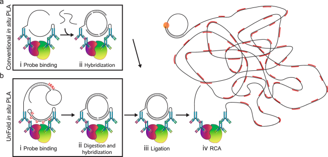
We have redesigned probes for in situ proximity ligation assay (PLA), resulting in more efficient localized detection of target proteins. In situ PLA depends on recognition of target proteins by pairs of antibody-oligonucleotide conjugates (PLA probes), which jointly give rise to DNA circles that template localized rolling circle amplification reactions. The requirement for dual recognition of the target proteins improves selectivity by ignoring any cross-reactivity not shared by the antibodies, and it allows detection of protein-protein interactions and post-translational modifications. We herein describe an improved design of the PLA probes -UnFold probes - where all elements required for formation of circular DNA strands are incorporated in the probes. Premature interactions between the UnFold probes are prevented by including an enzymatic "unfolding" step in the detection reactions. This allows DNA circles to form by pairs of reagents only after excess reagents have been removed. We demonstrate the performance of UnFold probes for detection of protein-protein interactions and post-translational modifications in fixed cells and tissues, revealing considerably more efficient signal generation. We also apply the UnFold probes to detect IL-6 in solution phase after capture on solid supports, demonstrating increased sensitivity over both normal sandwich enzyme-linked immunosorbent assays and conventional PLA assays.


Conflict of interest statement

U.L. is a founder and shareholder of Olink Biosciences having rights to the PLA technology. O.S. and U.L. are inventors of a patent covering the UnFold probe invention. A.K., K.G., T.E., J.H., B.K., M.L., D.R., J.O. and L.A. declare no competing interests.

Figures

Figure 1
Figure 1
Schematic illustration of in situ PLA using conventional and UnFold probes. (a) Conventional in situ PLA. (b) In situ PLA using UnFold probes. (i) After pairs of primary antibodies have bound a pair of interacting proteins (red and green) followed by washes, secondary conventional or UnFold in situ PLA probes are added, followed after an incubation by renewed washes. (ii) In the conventional design under (a) two more oligonucleotides are then added that can form a DNA circle. Using the UnFold design in (b) the probe carrying a hairpin-loop oligonucleotide is cleaved at the U residues, liberating a free 5′ end capable of being ligated to the 3′ end of the same DNA strand. Meanwhile, the U residues in the hairpin DNA strand of the other UnFold probe are cleaved presenting a single-stranded template for the enzymatic joining of the ends of the strand on the first UnFold probe. (iii) A DNA ligase is added to form DNA circles in the two variants of in situ PLA. (iv) Finally, phi29 DNA polymerase is added to initiate RCA primed by oligonucleotides on one of the antibodies, and fluorescent oligonucleotides are used to visualize the RCA products.
Figure 2
Figure 2
Visualization of E-cadherin/β-catenin interactions in HaCat cells. (a) Images of cells stained with primary antibodies against E-cadherin and β-catenin and secondary conventional or UnFold in situ PLA probes recording E-cadherin/β-catenin interactions (Pos). As a background control, primary antibodies were omitted (Neg). The RCA products were labeled with Cy3 (red dots in the merged images) and the nuclei were stained with Hoechst 33342. Scale bar (white) = 50 µm. (b) Quantification of signals per cell comparing in situ PLA and UnFold at different probe concentrations. Experiments were performed three times. Three images per experimental condition were acquired. Error bars represent SEM.
Figure 3
Figure 3
Image panel of HaCat cells showing detection of β-catenin/ E-cadherin interactions along with several technical controls. In the top row images, both of the primary antibodies and the PLA probes were included, revealing the characteristic β-catenin/ E-cadherin interaction stain. For the remaining rows in the panel, one or more of the primary or secondary antibodies were excluded. All signals were stained with Cy3 (Red in merge images and in grayscale) and the nuclei were stained with Hoechst 33342 (Blue). Scale bar (white) = 50 μm.
Figure 4
Figure 4
Image panel of HaCat cells showing detection of β-catenin/E-cadherin interaction or different technical controls, omitting enzymes. For the top image row, both of the primary antibodies and the PLA probes were included, revealing the characteristic staining pattern of β-catenin/E-cadherin interactions. The remaining rows in the panel show the effect of removing enzymes or detection oligonucleotides as indicated in the figure. All signals were stained with Cy3 (Red in merge images or in grayscale) and the nuclei were stained with Hoechst 33342 (Blue). Scale bar (white) = 50 μm.
Figure 5
Figure 5
Visualization of the interaction of β-catenin and E-cadherin in skin tissue. This stitched tile scan of stained skin tissue shows the specificity of the β-catenin and E-cadherin interaction (red) in the epidermis. The autofluorescence in the FITC channel (green), together with the nuclear Hoechst staining (blue) allows visualization of the different structures in the tissues. Each tissue section was stained with conventional or UnFold in situ PLA probes at a concentration of 1800 ng/ml and 66 ng/ml, respectively. Scale bar (white) = 100 µm. This experiment was performed once.
Figure 6
Figure 6
Visualization of phosphorylation of PDGFR-β in BJ hTert cells. (a) Images comparing serum-starved BJ hTert cells (−) versus cells treated with PDGF-BB for 45 min on ice (+) by detecting phosphorylation levels of the PDGFR-β (anti-PDGFR and anti-pan phosphorylation (pY100) antibodies). The RCA products were labeled with Cy3 (red dots in the merged images) and the nuclei were stained with Hoechst 33342. Scale bar (white) = 50 µm. (b) Quantification of signals comparing conventional and UnFold in situ PLA at different probe concentrations. The experiment was performed four times. Five images were collected per experimental condition. Error bars represent SEM.
Figure 7
Figure 7
Detection of recombinant IL-6 from solution phase using UnFold probes. Microtiter wells were precoated with capture antibodies, which served to bind the antigen, IL-6. Captured target proteins were then detected using a standard sandwich enzyme-linked immunosorbent assay (ELISA), or via conventional or UnFold in situ PLA probes. In each case, the signals were recorded by an HRP-mediated colorimetric reaction. After addition of the HRP substrate, TMB, the absorbance was recorded by spectrophotometry. The experiment was performed four times and one representative graph is shown. Error bars = Standard deviation of duplicates, dashed lines indicate the experimental LODs.

Similar articles

Cited by

References

    1. Wide L, Bennich H, Johansson SG. Diagnosis of allergy by an in-vitro test for allergen antibodies. Lancet. 1967;2:1105–1107. doi: 10.1016/S0140-6736(67)90615-0. - DOI - PubMed
    1. Coons A, Creech H, Jones RN, Berliner E. Demonstration of pneumoccocal antigen in tissues by use of fluorescent antibody. J Immunol. 1942;45:12.
    1. Raykova D, et al. Let There Be Light! Proteomes. 2016;4:36. doi: 10.3390/proteomes4040036. - DOI - PMC - PubMed
    1. Andersson, S. et al. Insufficient antibody validation challenges estrogen receptor beta research. Nature communications (2017). - PMC - PubMed
    1. Soderberg O, et al. Direct observation of individual endogenous protein complexes in situ by proximity ligation. Nature methods. 2006;3:995–1000. doi: 10.1038/nmeth947. - DOI - PubMed

Publication types

MeSH terms