

Genome-wide analysis of replication timing by next-generation sequencing with E/L Repli-seq
- PMID: 29599440
- PMCID: PMC6044726
- DOI: 10.1038/nprot.2017.148
Genome-wide analysis of replication timing by next-generation sequencing with E/L Repli-seq
Abstract
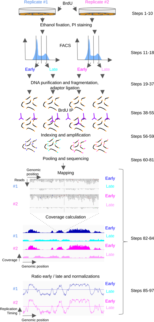
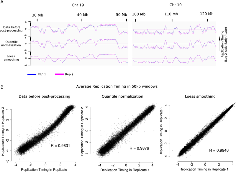
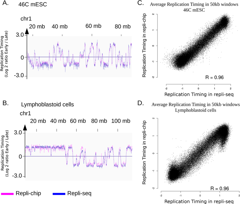
This protocol is an extension to: Nat. Protoc. 6, 870-895 (2014); doi:10.1038/nprot.2011.328; published online 02 June 2011Cycling cells duplicate their DNA content during S phase, following a defined program called replication timing (RT). Early- and late-replicating regions differ in terms of mutation rates, transcriptional activity, chromatin marks and subnuclear position. Moreover, RT is regulated during development and is altered in diseases. Here, we describe E/L Repli-seq, an extension of our Repli-chip protocol. E/L Repli-seq is a rapid, robust and relatively inexpensive protocol for analyzing RT by next-generation sequencing (NGS), allowing genome-wide assessment of how cellular processes are linked to RT. Briefly, cells are pulse-labeled with BrdU, and early and late S-phase fractions are sorted by flow cytometry. Labeled nascent DNA is immunoprecipitated from both fractions and sequenced. Data processing leads to a single bedGraph file containing the ratio of nascent DNA from early versus late S-phase fractions. The results are comparable to those of Repli-chip, with the additional benefits of genome-wide sequence information and an increased dynamic range. We also provide computational pipelines for downstream analyses, for parsing phased genomes using single-nucleotide polymorphisms (SNPs) to analyze RT allelic asynchrony, and for direct comparison to Repli-chip data. This protocol can be performed in up to 3 d before sequencing, and requires basic cellular and molecular biology skills, as well as a basic understanding of Unix and R.
Conflict of interest statement
COMPETING FINANCIAL INTERESTS
The authors declare that they have no competing financial interests.
Figures

Similar articles
-
Mapping Replication Timing in Single Mammalian Cells.Curr Protoc. 2022 Jan;2(1):e334. doi: 10.1002/cpz1.334. Curr Protoc. 2022. PMID: 34986273 Free PMC article.
-
Profiling Chromatin Accessibility on Replicated DNA with repli-ATAC-Seq.Methods Mol Biol. 2023;2611:71-84. doi: 10.1007/978-1-0716-2899-7_6. Methods Mol Biol. 2023. PMID: 36807065
-
Allele-specific control of replication timing and genome organization during development.Genome Res. 2018 Jun;28(6):800-811. doi: 10.1101/gr.232561.117. Epub 2018 May 7. Genome Res. 2018. PMID: 29735606 Free PMC article.
-
Genome Wide Approaches to Identify Protein-DNA Interactions.Curr Med Chem. 2019;26(42):7641-7654. doi: 10.2174/0929867325666180530115711. Curr Med Chem. 2019. PMID: 29848263 Review.
-
[The application of next generation sequencing on epigenetic study].Yi Chuan. 2014 Mar;36(3):256-75. Yi Chuan. 2014. PMID: 24846966 Review. Chinese.
Cited by
-
Rif1-Dependent Control of Replication Timing.Genes (Basel). 2022 Mar 20;13(3):550. doi: 10.3390/genes13030550. Genes (Basel). 2022. PMID: 35328102 Free PMC article. Review.
-
Diverse silent chromatin states modulate genome compartmentalization and loop extrusion barriers.Nat Struct Mol Biol. 2023 Jan;30(1):38-51. doi: 10.1038/s41594-022-00892-7. Epub 2022 Dec 22. Nat Struct Mol Biol. 2023. PMID: 36550219 Free PMC article.
-
Learning representations of chromatin contacts using a recurrent neural network identifies genomic drivers of conformation.Nat Commun. 2022 Jun 28;13(1):3704. doi: 10.1038/s41467-022-31337-w. Nat Commun. 2022. PMID: 35764630 Free PMC article.
-
Optimized Repli-seq: improved DNA replication timing analysis by next-generation sequencing.Chromosome Res. 2022 Dec;30(4):401-414. doi: 10.1007/s10577-022-09703-7. Epub 2022 Jul 4. Chromosome Res. 2022. PMID: 35781769 Free PMC article.
-
DNA Replication Timing Enters the Single-Cell Era.Genes (Basel). 2019 Mar 15;10(3):221. doi: 10.3390/genes10030221. Genes (Basel). 2019. PMID: 30884743 Free PMC article. Review.
References
KEY REFERENCES:
Publication types
MeSH terms
Substances
Grants and funding
LinkOut - more resources
Full Text Sources
Other Literature Sources
Molecular Biology Databases
