Review
. 2018 Jun;16(6):355-367.
doi: 10.1038/s41579-018-0001-8.

Streptococcus pneumoniae: transmission, colonization and invasion

Affiliations
Review

Streptococcus pneumoniae: transmission, colonization and invasion

Jeffrey N Weiser et al. Nat Rev Microbiol. 2018 Jun.

Abstract

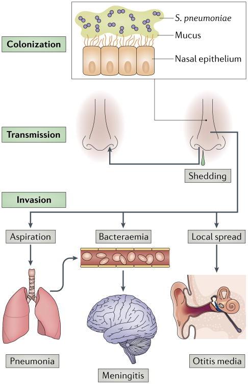
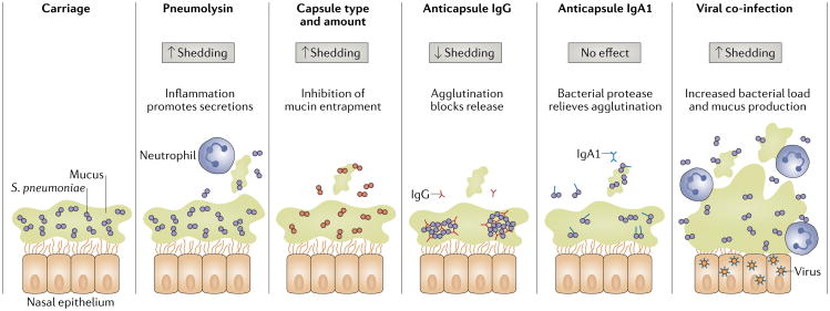
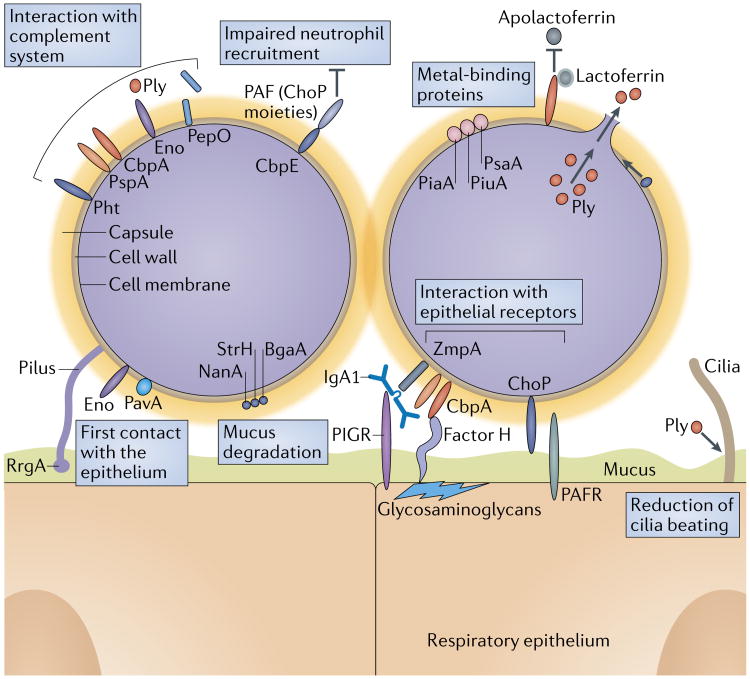
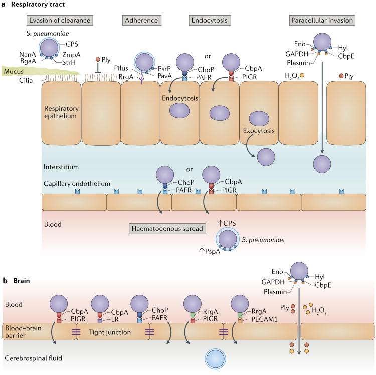
Streptococcus pneumoniae has a complex relationship with its obligate human host. On the one hand, the pneumococci are highly adapted commensals, and their main reservoir on the mucosal surface of the upper airways of carriers enables transmission. On the other hand, they can cause severe disease when bacterial and host factors allow them to invade essentially sterile sites, such as the middle ear spaces, lungs, bloodstream and meninges. Transmission, colonization and invasion depend on the remarkable ability of S. pneumoniae to evade or take advantage of the host inflammatory and immune responses. The different stages of pneumococcal carriage and disease have been investigated in detail in animal models and, more recently, in experimental human infection. Furthermore, widespread vaccination and the resulting immune pressure have shed light on pneumococcal population dynamics and pathogenesis. Here, we review the mechanistic insights provided by these studies on the multiple and varied interactions of the pneumococcus and its host.


Figures

Fig. 1
Fig. 1. The life cycle of Streptococcus pneumoniae and the pathogenesis of pneumococcal disease
Streptococcus pneumoniae colonizes the mucosa of the upper respiratory tract (URT). This carriage is the prerequisite for both transmission to other individuals and invasive disease in the carrier. Carriers can shed S. pneumoniae in nasal secretions and thereby transmit the bacterium. Dissemination beyond its niche along the nasal epithelium, either by aspiration, bacteraemia or local spread, can lead to invasive diseases, such as pneumonia, meningitis and otitis media.
Fig. 2
Fig. 2. bacterial and host factors affecting pneumococcal shedding from carriers
Streptococcus pneumoniae is found predominantly in the mucus layer overlying the epithelial surface of the upper respiratory tract. Inflammation (indicated by the presence of neutrophils), which is induced by the pore-forming toxin pneumolysin or by co-infection with influenza virus or other respiratory viruses, stimulates secretions and increases shedding. By contrast, agglutinating antibodies such as anti-capsule immunoglobulin G (IgG) and IgA1 decrease shedding unless they are cleaved by the human IgA1-specific pneumococcal protease. Capsule type and amount also influence mucus association and numbers of shed bacteria.
Fig. 3
Fig. 3. Molecular mechanisms of pneumococcal colonization of host surfaces
Key functions that enable Streptococcus pneumoniae colonization are: establishing the first contact with the epithelium and epithelial receptors, interaction with the complement system, mucus degradation, metal binding, impairment of neutrophil activity and the pro-inflammatory effects of the toxin pneumolysin (Ply). The pneumococcal enzymes Neuraminidase A (NanA), β-galactosidase (BgaA) and β-N-acetylglucosaminidase (StrH) degrade mucus and thereby inhibit mucociliary clearance. Furthermore, the LytA (autolysin)-facilitated release of Ply damages the epithelium and reduces ciliary beating. Negatively charged capsular polysaccharide (CPS) inhibits bacterial mucus entrapment. CPS and several pneumococcal proteins, including pneumococcal surface protein A (PspA), choline-binding protein A (CbpA), enolase (Eno) and pneumococcal histidine triad protein (Pht), directly and indirectly block complement deposition. PspA also binds to lactoferrin to acquire iron and blocks the antimicrobial effect of apolactoferrin. Endopeptidase (PepO), which is released from the pneumococcal surface, binds to C1q and thereby depletes complement components. Pneumococcal CbpE impairs neutrophil recruitment by degrading platelet-activating factor (PAF), a host-derived inflammatory phospholipid. CbpA interacts with factor H interactions to facilitate adherence and subsequent internalization of S. pneumoniae via cell glycosaminoglycans. CbpA also binds to polymeric immunoglobulin receptor (PIGR) to promote adherence. The zinc metalloprotease ZmpA (also known as immunoglobulin A1 protease) subverts mucosal humoral immunity by cleaving IgA1. Phosphorylcholine (ChoP) on teichoic acid mimics host PAF and allows binding to its receptor. Piliated strains express an ancillary pilus subunit tip adhesin called RrgA. Other S. pneumoniae adhesins include enolase (Eno) and adherence and virulence protein A (PavA). PAFR, platelet-activating factor receptor.
Fig. 4
Fig. 4. Stages in pneumococcal adherence and invasion
a | Several steps are required for invasion of the respiratory tract. Streptococcus pneumoniae evades entrapment in mucus and mucociliary clearance by negatively charged capsular polysaccharide (CPS) and proteolytic degradation of secretory immunoglobulin A1 (IgA1) by the zinc metalloprotease ZmpA (also known as IgA1 protease). Neuraminidase A (NanA), β-galactosidase (BgaA) and β-N-acetylglucosaminidase (StrH) deglycosylate mucus and unmask glycan targets for adhesins on the epithelium. Finally, pneumolysin (Ply) inhibits ciliary beating. Adherence to the apical surface of epithelial cells is mediated by diverse surface structures, including phosphorylcholine (ChoP), choline-binding protein A (CbpA), the ancillary pilus subunit RrgA at the tip of pili, adherence and virulence protein A (PavA) and large surface-exposed glycoprotein (PsrP). S. pneumoniae binds through ChoP to platelet-activating factor receptor (PAFR) and through CbpA to polymeric immunoglobulin receptor (PIGR), and by subverting the respective host receptor recycling pathways, it induces its own endocytosis, which is followed by release of pneumococci at the basolateral surface. Alternatively, Ply and hydrogen peroxide (H2O2) directly damage the epithelium, and hyaluronate lyase (Hyl) and plasmin, which is bound to the pneumococcal surface through enolase (Eno), glyceralde-hyde-3-phosphate dehydrogenase (GAPDH) or CbpE, degrade the extracellular matrix. This breaks down the epithelial barrier and provides a pathway for paracellular invasion. ChoP–PAFR and CbpA–PIGR interactions also enable pneumococci to traverse the endothelium and enter the bloodstream. Upregulation of PAFR by inflammatory cytokines amplifies ChoP–PAFR-mediated invasion. CPS and other virulence factors, including pneumococcal surface protein A (PspA), CbpA and Ply, facilitate evasion of opsonophagocytosis. b | To penetrate the blood–brain barrier, S. pneumoniae uses ChoP–PAFR, CbpA–PIGR and CbpA–laminin receptor (LR) binding. Strains that express pili also use RrgA to bind to PIGR and platelet endothelial cell adhesion molecule 1 (PECAM1). Similar to invasion of the respiratory tract, Ply, H2O2 generated by α-glycerophosphate oxidase (GlpO) and activated plasmin bound to the pneumococcal surface proteins Eno, GAPDH and CbpE can compromise the blood–brain barrier.

References

    1. Abdullahi O, et al. The prevalence and risk factors for pneumococcal colonization of the nasopharynx among children in Kilifi District, Kenya. PLoS ONE. 2012;7:e30787. - PMC - PubMed
    1. Yahiaoui RY, et al. Prevalence and antibiotic resistance of commensal Streptococcus pneumoniae in nine European countries. Future Microbiol. 2016;11:737–744. - PubMed
    1. Bogaert D, De Groot R, Hermans PW. Streptococcus pneumoniae colonisation: the key to pneumococcal disease. Lancet Infect Dis. 2004;4:144–154. - PubMed
    1. Whitney CG, et al. Decline in invasive pneumococcal disease after the introduction of protein-polysaccharide conjugate vaccine. N Engl J Med. 2003;348:1737–1746. - PubMed
    1. Musher D. How contagious are common respiratory tract infections? N Engl J Med. 2003;348:1256–1266. - PubMed

Publication types

MeSH terms