

Activation of Eosinophils and Mast Cells in Functional Dyspepsia: an Ultrastructural Evaluation
- PMID: 29599471
- PMCID: PMC5876347
- DOI: 10.1038/s41598-018-23620-y
Activation of Eosinophils and Mast Cells in Functional Dyspepsia: an Ultrastructural Evaluation
Abstract
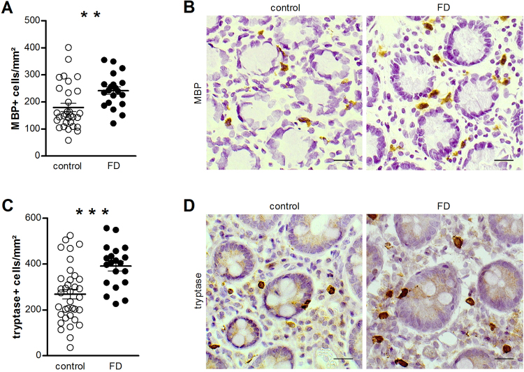
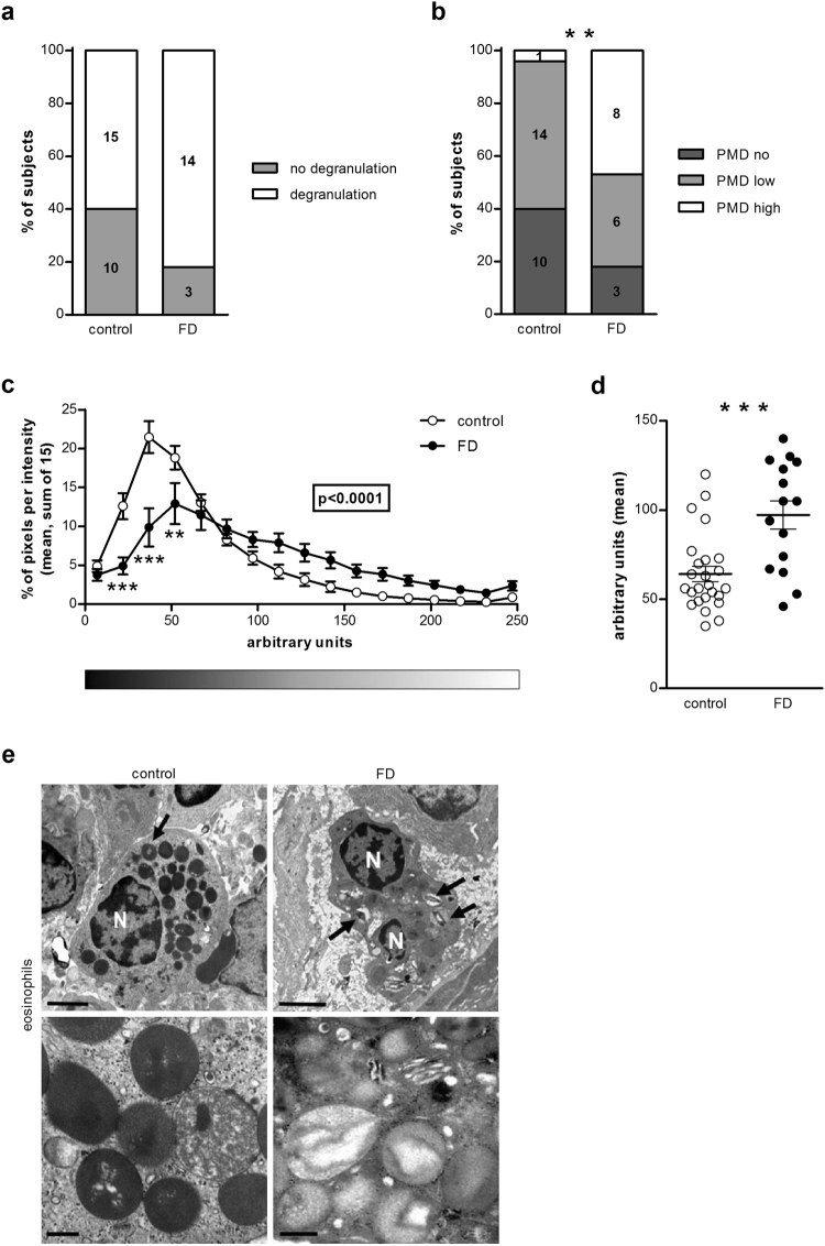
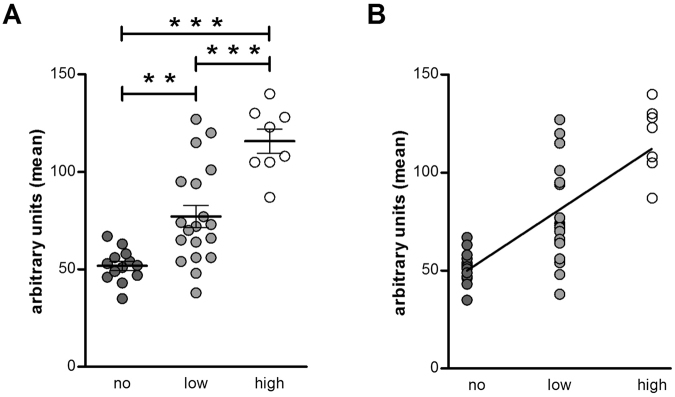
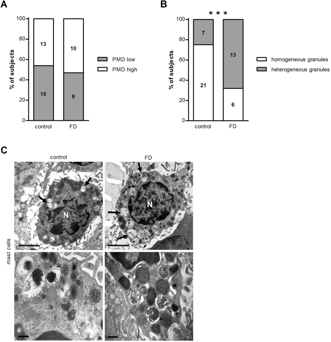
We recently identified mucosal mast cell and eosinophil hyperplasia in association with a duodenal impaired barrier function in functional dyspepsia (FD). We aimed to further describe the implication of these immune cells by assessing their activation state at the ultrastructural level and by evaluating the association between impaired epithelial integrity and immune activation. Duodenal biopsies were obtained from 24 FD patients and 37 healthy controls. The ultrastructure of mast cells and eosinophils was analyzed by transmission electron microscopy. Transepithelial electrical resistance and paracellular permeability were measured to evaluate epithelial barrier function. The type of degranulation in eosinophils and mast cells was piecemeal. Eosinophils displayed higher degree of degranulation in FD patients than in controls (p < 0.0001). Quantification revealed a decreased granular density in eosinophils of FD patients (p < 0.0001). The degree of degranulation in mast cells was similar in both groups. However, a more heterogeneous profile was found in the FD group (p < 0.0001). No association between epithelial integrity and the number and activation state of mucosal eosinophils and mast cells was found. We demonstrated ultrastructural changes in degranulation state of eosinophils and mast cells, suggesting that eosinophil and mast cell activation play a role in the pathophysiology of FD.
Conflict of interest statement
The authors declare no competing interests.
Figures
Similar articles
-
Quantitative evaluation of duodenal eosinophils and mast cells in adult patients with functional dyspepsia.Ann Diagn Pathol. 2015 Apr;19(2):50-6. doi: 10.1016/j.anndiagpath.2015.02.001. Epub 2015 Feb 20. Ann Diagn Pathol. 2015. PMID: 25735567
-
Increased counts and degranulation of duodenal mast cells and eosinophils in functional dyspepsia- a clinical study.Med Glas (Zenica). 2014 Aug;11(2):276-82. Med Glas (Zenica). 2014. Retraction in: Med Glas (Zenica). 2015 Feb;12(1):107. PMID: 25082240 Retracted.
-
Increased Duodenal Eosinophil Degranulation in Patients with Functional Dyspepsia: A Prospective Study.Sci Rep. 2016 Oct 6;6:34305. doi: 10.1038/srep34305. Sci Rep. 2016. PMID: 27708358 Free PMC article. Clinical Trial.
-
Micro-inflammation in functional dyspepsia: A systematic review and meta-analysis.Neurogastroenterol Motil. 2018 Apr;30(4):e13304. doi: 10.1111/nmo.13304. Epub 2018 Feb 2. Neurogastroenterol Motil. 2018. PMID: 29392796
-
Beyond Eosinophilic Esophagitis: Eosinophils in Gastrointestinal Disease-New Insights, "New" Diseases.J Can Assoc Gastroenterol. 2023 Nov 24;6(6):199-211. doi: 10.1093/jcag/gwad046. eCollection 2023 Dec. J Can Assoc Gastroenterol. 2023. PMID: 38106480 Free PMC article. Review.
Cited by
-
New Insights into Intestinal Permeability in Irritable Bowel Syndrome-Like Disorders: Histological and Ultrastructural Findings of Duodenal Biopsies.Cells. 2021 Sep 29;10(10):2593. doi: 10.3390/cells10102593. Cells. 2021. PMID: 34685576 Free PMC article.
-
A Pilot Study of Ketotifen in Patients Aged 8-17 Years with Functional Dyspepsia Associated with Mucosal Eosinophilia.Paediatr Drugs. 2024 Jul;26(4):451-457. doi: 10.1007/s40272-024-00628-8. Epub 2024 May 21. Paediatr Drugs. 2024. PMID: 38771467 Clinical Trial.
-
Associations among the Duodenal Ecosystem, Gut Microbiota, and Nutrient Intake in Functional Dyspepsia.Gut Liver. 2024 Jul 15;18(4):621-631. doi: 10.5009/gnl230130. Epub 2023 Nov 30. Gut Liver. 2024. PMID: 38031491 Free PMC article.
-
Potential neuro-immune therapeutic targets in irritable bowel syndrome.Therap Adv Gastroenterol. 2020 Apr 9;13:1756284820910630. doi: 10.1177/1756284820910630. eCollection 2020. Therap Adv Gastroenterol. 2020. PMID: 32313554 Free PMC article. Review.
-
Intestinal Mucosal Mast Cells: Key Modulators of Barrier Function and Homeostasis.Cells. 2019 Feb 8;8(2):135. doi: 10.3390/cells8020135. Cells. 2019. PMID: 30744042 Free PMC article. Review.
References
-
- Stanghellini V, et al. Rome IV - Gastroduodenal Disorders. Gastroenterology. 2016 - PubMed
Publication types
MeSH terms
LinkOut - more resources
Full Text Sources
Other Literature Sources
Medical
