

Corneal Innervation and Sensation: The Eye and Beyond
- PMID: 29599653
- PMCID: PMC5872636
Corneal Innervation and Sensation: The Eye and Beyond
Abstract
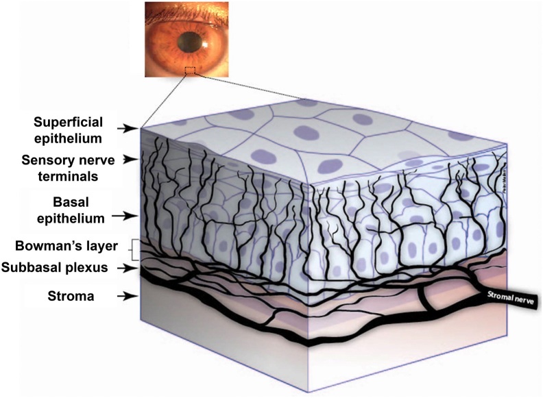
The cornea is one of the most densely innervated and sensitive tissues in the body. In addition to their important sensory functions, corneal nerves induce reflex tear production, blinking, and the release of trophic factors - all of which combined help to maintain the structural and functional integrity of the surface of the eye. Consequently, damage to corneal nerves as a result of disease, surgery, or trauma can lead to diminished corneal sensitivity, epithelial defects, and possible blindness. In this review, we describe commonly used tools that have provided considerable new information on corneal architecture and sensation in healthy and diseased corneas, with special emphasis on changes seen in herpes zoster ophthalmicus, corneal and other therapeutic ocular procedures, antiglaucoma medical therapy, aging, and diabetes. With its potential applications ranging from managing ocular-specific to systemic diseases, the study of corneal innervation has implications for future therapies extending beyond just the eye itself.
Keywords: corneal innervation; corneal sensation; ophthalmic disease; ophthalmology; subbasal nerve plexus.
Figures
References
-
- Bonini S, Rama P, Olzi D, et al. Neurotrophic keratitis. Eye (Lond). 2003;17:989–95. - PubMed
-
- Kitano S. An embryologic study of the human corneal nerves. Jpn J Ophthalmol. 1957;1:48–55.
-
- Marfurt CF, Kingsley RE, Echtenkamp SE. Sensory and sympathetic innervation of the mammalian cornea. A retrograde tracing study. Invest Ophthalmol Vis Sci. 1989;30(3):461–72. - PubMed
-
- Müller LJ, Marfurt CF, Kruse F, Tervo TM. Corneal nerves: structure, contents and function. Exp Eye Res. 2003;76(5):521–42. - PubMed
Publication types
MeSH terms
LinkOut - more resources
Full Text Sources