Review
. 2018 Feb;35(1):6-17.
doi: 10.5114/pdia.2017.67095. Epub 2018 Feb 20.

Langerhans cell histiocytosis in children - a disease with many faces. Recent advances in pathogenesis, diagnostic examinations and treatment

Affiliations
Review

Langerhans cell histiocytosis in children - a disease with many faces. Recent advances in pathogenesis, diagnostic examinations and treatment

Michalina Jezierska et al. Postepy Dermatol Alergol. 2018 Feb.

Abstract

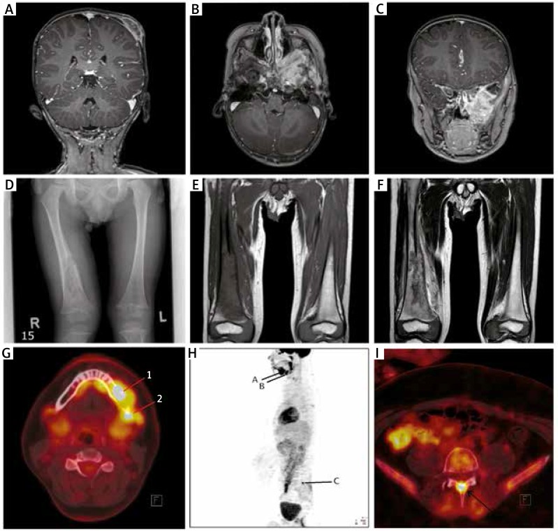
Langerhans cell histiocytosis is a rare clonal disease characterized by the proliferation of CD1a-positive immature dendritic cells. The purpose of this article was to present an updated review of recent advances in the pathogenesis, clinical features, imaging and treatment of this disease. The discovery of oncogenic BRAF mutations and the presence of proinflammatory cytokines and chemokines confirmed the unusual characteristics of this disease. Currently, children with organ involvement who do not have a good response to chemotherapy and have neurodegeneration or diabetes insipidus are the most problematic patients. Further research is needed to improve the results of treatment.

Keywords: Langerhans cell histiocytosis; children; pathogenesis; symptoms; treatment.


Figures

Figure 1
Figure 1
A – Magnetic resonance imaging (T1-weighted image) after contrast administration – osteolytic lesion of the parietal bone with soft tissue involvement. B – Magnetic resonance imaging (T1-weighted image) after contrast administration – extensive infiltration of the skull base (left side), horizontal projection. C – Magnetic resonance imaging (T1-weighted image) after contrast administration – extensive infiltration of the skull base (left side), vertical projection. D – X-ray, PA – osteolytic focus of the right femur with interruption of cortical layer and periosteal reaction. E – Magnetic resonance imaging (T1-weighted image) in the frontal plane – destruction of the distal shaft of the right femur. F – Magnetic resonance imaging (T2-weighted image) in the frontal plane – destruction of the distal shaft of the right femur. G – PET-CT FDG and CT fusion image – hypermetabolic mass in the left side mandible with the bone lesion (SUVmax 15.9) (1) and involved submandibular hypermetabolic lymph nodes (SUVmax 11.3) (2). H – WB FDG-PET scan in lateral projection showing hypermetabolic activity on the left side of the mandible (A) and adjacent lymph nodes (B), as well as in the arch of L5 (C). I – FDG-PET and CT fusion image with a hypermetabolic mass in the arch of L5, which resulted in a bone lesion (SUVmax 11.4)
Figure 2
Figure 2
A – Ultrasonography (1HX1 elastography) of the intraosteal cyst of the temporal bone with no visible features of bone destruction. B – Ultrasonography (1HX1 elastography) of a solid lesion in the right parietal area with bone destruction. The blue colour corresponds to a hard lesion, and the image suggests a proliferative process. C – Computed tomography (CT) of the abdomen – hepatomegaly. D – Computed tomography (CT) of the abdomen – splenomegaly

Similar articles

Cited by

References

    1. Emile JF, Abla O, Fraitag S, et al. Revised classification of histiocytoses and neoplasms of the macrophage-dendritic cell lineages. Blood. 2016;127:2672–81. - PMC - PubMed
    1. Haupt R, Minkov M, Astigarraga I, et al. Langerhans cell histiocytosis (LCH): guidelines for diagnosis, clinical work-up, and treatment for patient till age of 18 years. Pediatr Blood Cancer. 2013;60:175–84. - PMC - PubMed
    1. Minkov M. Multisystem Langerhans cell histiocytosis in children. Current treatment and future directions. Pediatr Drugs. 2011;13:75–86. - PubMed
    1. Margo CE, Goldman DR. Langerhans cell histiocytosis. Surv Ophthalmol. 2008;53:332–58. - PubMed
    1. Grana N. Langerhans cell histiocytosis. Cancer Control. 2014;21:328–34. - PubMed