

Caffeine and Modafinil Ameliorate the Neuroinflammation and Anxious Behavior in Rats during Sleep Deprivation by Inhibiting the Microglia Activation
- PMID: 29599709
- PMCID: PMC5863523
- DOI: 10.3389/fncel.2018.00049
Caffeine and Modafinil Ameliorate the Neuroinflammation and Anxious Behavior in Rats during Sleep Deprivation by Inhibiting the Microglia Activation
Abstract
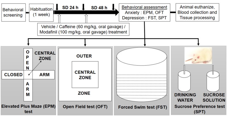
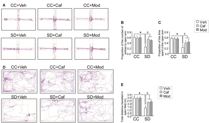
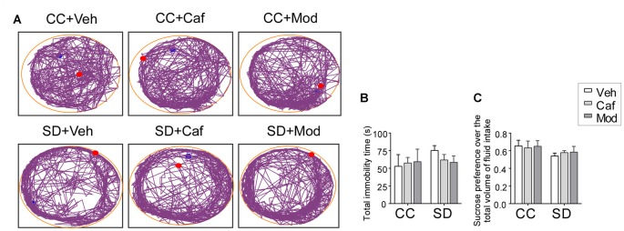
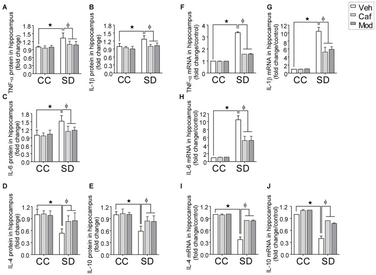
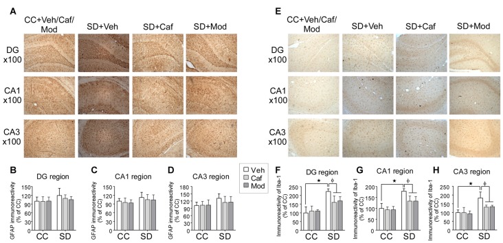
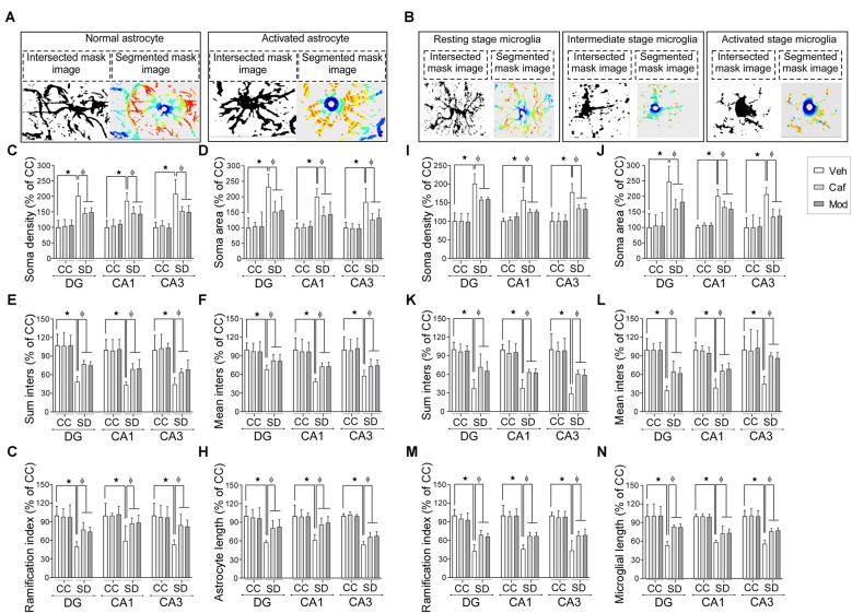
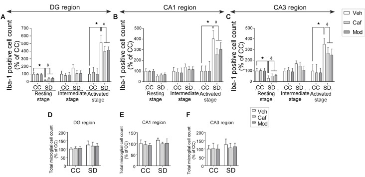
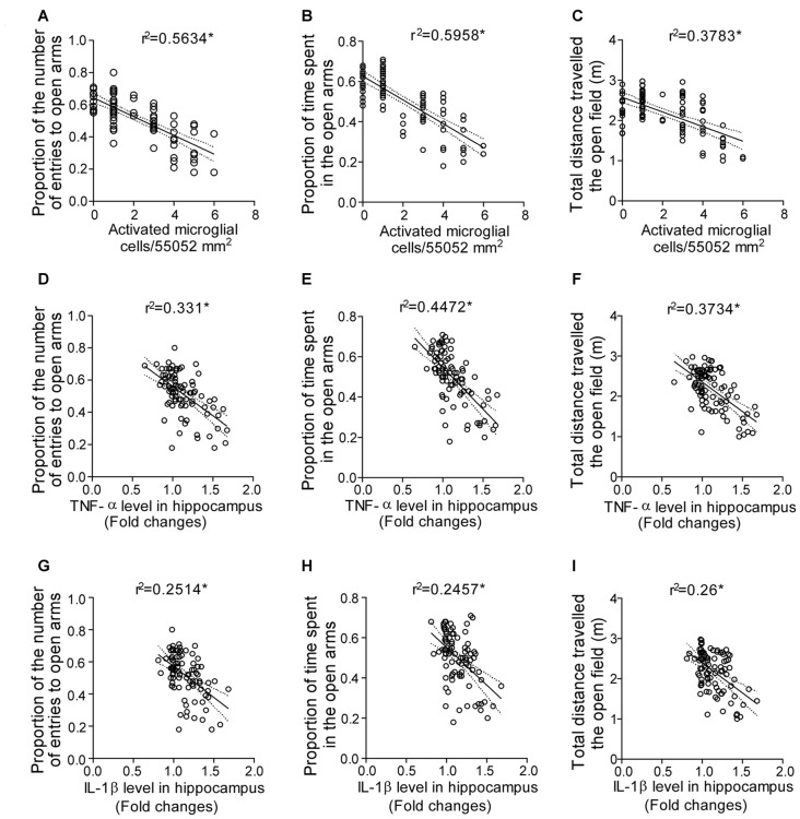
Background: Sleep deprivation (SD) plagues modern society due to the professional demands. It prevails in patients with mood and neuroinflammatory disorders. Although growing evidence suggests the improvement in the cognitive performance by psychostimulants during sleep-deprived conditions, the impending involved mechanism is rarely studied. Thus, we hypothesized that mood and inflammatory changes might be due to the glial cells activation induced modulation of the inflammatory cytokines during SD, which could be improved by administering psychostimulants. The present study evaluated the role of caffeine/modafinil on SD-induced behavioral and inflammatory consequences. Methods: Adult male Sprague-Dawley rats were sleep deprived for 48 h using automated SD apparatus. Caffeine (60 mg/kg/day) or modafinil (100 mg/kg/day) were administered orally to rats once every day during SD. Rats were subjected to anxious and depressive behavioral evaluation after SD. Subsequently, blood and brain were collected for biochemical, immunohistochemical and molecular studies. Results: Sleep deprived rats presented an increased number of entries and time spent in closed arms in elevated plus maze test and decreased total distance traveled in the open field (OF) test. Caffeine/modafinil treatment significantly improved these anxious consequences. However, we did not observe substantial changes in immobility and anhedonia in sleep-deprived rats. Caffeine/modafinil significantly down-regulated the pro- and up-regulated the anti-inflammatory cytokine mRNA and protein expression in the hippocampus during SD. Similar outcomes were observed in blood plasma cytokine levels. Caffeine/modafinil treatment significantly decreased the microglial immunoreactivity in DG, CA1 and CA3 regions of the hippocampus during SD, however, no significant increase in immunoreactivity of astrocytes was observed. Sholl analysis signified the improvement in the morphological alterations of astrocytes and microglia after caffeine/modafinil administration during SD. Stereological analysis demonstrated a significant improvement in the number of ionized calcium binding adapter molecule I (Iba-1) positive cells (different states) in different regions of the hippocampus after caffeine or modafinil treatment during SD without showing any significant change in total microglial cell number. Eventually, the correlation analysis displayed a positive relationship between anxiety, pro-inflammatory cytokines and activated microglial cell count during SD. Conclusion: The present study suggests the role of caffeine or modafinil in the amelioration of SD-induced inflammatory response and anxious behavior in rats. Highlights - SD induced mood alterations in rats. - Glial cells activated in association with the changes in the inflammatory cytokines. - Caffeine or modafinil improved the mood and restored inflammatory changes during SD. - SD-induced anxious behavior correlated with the inflammatory consequences.
Keywords: caffeine; cytokines; microglia; modafinil; mood changes; neuroinflammation; sleep deprivation.
Figures
Similar articles
-
Caffeine and modafinil promote adult neuronal cell proliferation during 48 h of total sleep deprivation in rat dentate gyrus.Exp Neurol. 2013 Oct;248:470-81. doi: 10.1016/j.expneurol.2013.07.021. Epub 2013 Aug 3. Exp Neurol. 2013. PMID: 23920241
-
Inhibiting the microglia activation improves the spatial memory and adult neurogenesis in rat hippocampus during 48 h of sleep deprivation.J Neuroinflammation. 2017 Nov 15;14(1):222. doi: 10.1186/s12974-017-0998-z. J Neuroinflammation. 2017. Retraction in: J Neuroinflammation. 2024 Aug 17;21(1):204. doi: 10.1186/s12974-024-03200-w. PMID: 29141671 Free PMC article. Retracted.
-
Complement activation sustains neuroinflammation and deteriorates adult neurogenesis and spatial memory impairment in rat hippocampus following sleep deprivation.Brain Behav Immun. 2019 Nov;82:129-144. doi: 10.1016/j.bbi.2019.08.004. Epub 2019 Aug 10. Brain Behav Immun. 2019. PMID: 31408672
-
Modafinil--medical considerations for use in sustained operations.Aviat Space Environ Med. 2003 Jun;74(6 Pt 1):659-63. Aviat Space Environ Med. 2003. PMID: 12793539 Review.
-
Effects of modafinil on cognitive performance and alertness during sleep deprivation.Curr Pharm Des. 2006;12(20):2457-71. doi: 10.2174/138161206777698819. Curr Pharm Des. 2006. PMID: 16842170 Review.
Cited by
-
Alterations in microglial morphology concentrate in the habitual sleeping period of the mouse.Glia. 2023 Feb;71(2):366-376. doi: 10.1002/glia.24279. Epub 2022 Oct 5. Glia. 2023. PMID: 36196985 Free PMC article.
-
Vibration-reduced anxiety-like behavior relies on ameliorating abnormalities of the somatosensory cortex and medial prefrontal cortex.Neural Regen Res. 2024 Jun 1;19(6):1351-1359. doi: 10.4103/1673-5374.385840. Epub 2023 Sep 22. Neural Regen Res. 2024. PMID: 37905885 Free PMC article.
-
Naringin Regulates Microglia BV-2 Activation and Inflammation via the JAK/STAT3 Pathway.Evid Based Complement Alternat Med. 2022 May 19;2022:3492058. doi: 10.1155/2022/3492058. eCollection 2022. Evid Based Complement Alternat Med. 2022. PMID: 35646153 Free PMC article.
-
The Effects of Antioxidant Approved Drugs and Under Investigation Compounds with Potential of Improving Sleep Disorders and their Associated Comorbidities Associated with Oxidative Stress and Inflammation.Mini Rev Med Chem. 2025;25(10):795-815. doi: 10.2174/0113895575360959250117073046. Mini Rev Med Chem. 2025. PMID: 39957704 Review.
-
Differentially Expressed Genes in the Brain of Aging Mice With Cognitive Alteration and Depression- and Anxiety-Like Behaviors.Front Cell Dev Biol. 2020 Aug 28;8:814. doi: 10.3389/fcell.2020.00814. eCollection 2020. Front Cell Dev Biol. 2020. PMID: 33015035 Free PMC article.
References
-
- Antoniou K., Kafetzopoulos E., Papadopoulou-Daifoti Z., Hyphantis T., Marselos M. (1998). D-amphetamine, cocaine and caffeine: a comparative study of acute effects on locomotor activity and behavioural patterns in rats. Neurosci. Biobehav. Rev. 23, 189–196. 10.1016/s0149-7634(98)00020-7 - DOI - PubMed
LinkOut - more resources
Full Text Sources
Other Literature Sources
Research Materials
Miscellaneous