. 2018 Feb 27:9:320.
doi: 10.3389/fmicb.2018.00320. eCollection 2018.

Mesobuthus Venom-Derived Antimicrobial Peptides Possess Intrinsic Multifunctionality and Differential Potential as Drugs

Affiliations

Mesobuthus Venom-Derived Antimicrobial Peptides Possess Intrinsic Multifunctionality and Differential Potential as Drugs

Bin Gao et al. Front Microbiol. .

Abstract

Animal venoms are a mixture of peptides and proteins that serve two basic biological functions: predation and defense against both predators and microbes. Antimicrobial peptides (AMPs) are a common component extensively present in various scorpion venoms (herein abbreviated as svAMPs). However, their roles in predation and defense against predators and potential as drugs are poorly understood. Here, we report five new venom peptides with antimicrobial activity from two Mesobuthus scorpion species. These α-helical linear peptides displayed highly bactericidal activity toward all the Gram-positive bacteria used here but differential activity against Gram-negative bacteria and fungi. In addition to the antibiotic activity, these AMPs displayed lethality to houseflies and hemotoxin-like toxicity on mice by causing hemolysis, tissue damage and inducing inflammatory pain. Unlike AMPs from other origins, these venom-derived AMPs seem to be unsuitable as anti-infective drugs due to their high hemolysis and low serum stability. However, MeuTXKβ1, a known two-domain Mesobuthus AMP, is an exception since it exhibits high activity toward antibiotic resistant Staphylococci clinical isolates with low hemolysis and high serum stability. The findings that the classical AMPs play predatory and defensive roles indicate that the multifunctionality of scorpion venom components is an intrinsic feature likely evolved by natural selection from microbes, prey and predators of scorpions. This definitely provides an excellent system in which one can study how a protein adaptively evolves novel functions in a new environment. Meantimes, new strategies are needed to remove the toxicity of svAMPs on eukaryotic cells when they are used as leads for anti-infective drugs.

Keywords: Staphylococci; defense; peptide antibiotics; predation; venom gland immunity.


Figures

Figure 1
Figure 1
MeuFSPLs and homologs. For each peptide, scorpion species and peptide names are shown. Glycines for C-terminal amidation are italicized and boxed in cyan. PCS, proprotein convertase signal. The arrow represents scissile peptide bond by PC. In the mature region, residues non-identical to MeuFSPL-1 are shadowed in yellow, and in the proregion, charged residues are colored (basic, blue; acidic, red). “a,” amidation. Non-identical residues between MeuFSPL-1 and BmKb1 are underlined once. Except three from frogs (Phyllobates azzurea, P. bicolor, and P. jandaia), all other sequences are derived from scorpions (Androctonus amoreuxi, Isometrus maculatus, Lychas mucronatus, Mesobuthus eupeus, Mesobuthus martensii, Tityus costatus, and Tityus serrulatus). These sequences can be retrieved from GenBank. “” represents new sequences reported in this work. The proposed mature peptide encoded by BmKb1* (i.e., marmelittin) is boxed in orange and the premature stop codon is shown by “ξ” in red.
Figure 2
Figure 2
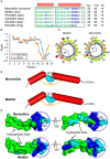
Marmelittin and Melittins. (A) Amino acid sequences. Hydrophobic and cationic residues are shown in green and blue, respectively. “a,” C-terminal amidation; α-helices, indicated by red cylinder, are extracted from the structural coordination of melittin (pdb 2MLT). The structural Gly is boxed in cyan. H (%), the percentage of hydrophobic amino acids. NC, net charge. The source of the sequences used here: Marmelittin from Mesobuthus martensii (AF159979), melittin from Apis mellifera (NP_001011607.1); Phmelittin from Polistes hebraeus (P59261); Admelittin from Apis dorsata (XP_006611828); Afmelittin from Apis florea (P01504); Rtmellitin from the frog Rana tagoi (BAF74741). (B) Hydropathy plots showing hydropathy scores for all the amino acids in the peptides. (C) Helical wheel projections of marmelittin and melittin. Hydrophilic, hydrophobic, negatively charged, and positively charged residues are presented as circles, diamonds, triangles, and pentagons, respectively. The most hydrophobic residue is shown in green, and the amount of green is decreasing proportionally to the hydrophobicity. The most hydrophilic (uncharged) residues are coded red, and the amount of red is decreasing proportionally to the hydrophilicity. Charged residues are shown in light blue. PF, polar face. (D) Structural similarity between marmelittin and melittin. The conserved Gly12 disrupting the helix is shown as cyan spheres. (E) Molecular surfaces of marmelittin and melittin with hydrophobic and hydrophilic regions indicated in green and cyan, respectively. The C-terminal charged/polar region is circled. The experimental structure of melittin (pdb 2MLT) was used as template to generate the structure of marmelittin. The method is detailed in Materials and Methods.
Figure 3
Figure 3
Polymorphism of BmKb1. (A) Precursor proteins encoded by BmKb1 and its mutants. Variations at site 50 are shown in red and identical sites are shadowed in yellow. (B) Sequencing PCR products amplified from genomic DNA and cDNA for identifying polymorphic sites. (C) Sequencing different genomic clones identifying polymorphism in the intron region of BmKb1.
Figure 4
Figure 4
Meucin-22 and homologs. (A) Precursor amino acid sequences. The green arrow indicates a newly proposed cleavage site based on the presence of a putative proprotein convertase signal (PCS), which leads to a C-terminal extension of four residues relative to the initially proposed Meucin-18, indicated by a black arrow. In the mature region, the amino acids non-identical to Meucin-22 are shadowed in yellow and in the proregion, charged residues are colored (basic, blue; acidic, red). The secondary structures of Meucin-22 and its homologs comprising an N-terminal amphipathic domain (residues 1–18, green and blue) and a charged tail (residues 19–22, red) are showed at the top of the sequence alignment. (B) Isolation of native Meucin-22, along with MeuTXKβ-1 from the venom of M. eupeus by RP-HPLC. Inset, MALDI-TOF MS of Meucin-22 and MeuTXKβ-1. For the spectrum of the latter, there are two main peaks, corresponding to the singly and doubly protonated forms of this peptide. (C) Helical wheel projection of Meucin-22. Color codes of amino acids are identical to those of Figure 2C. (D) Ribbon representation of Meucin-22 backbone structure that was built based on the experimental structure of racemic Ala-(8,13,18) Magainin 2 (pdb 4MGP), an analog of frog skin AMP. Residues with different side-chain natures are indicated in different colors (blue, positively charged; red, negatively charged; green, hydrophobic; cyan, polar) on the background of a semitransparent surface. In the helix formed by the N-terminal 18 residues, all hydrophobic residues are located on one surface while cationic and hydrophilic residues on another surface.The figure was prepared with PyMOL (http://www.pymol.org).
Figure 5
Figure 5
Purification of synthetic svAMPs by RP-HPLC. For each peptide, their sequences and retention times (TR) are shown. Pure peptides indicated by triangle were collected for structural and funtional studies. The leucine/isoleucine residues presumably forming a short leucine zipper-like motif are marked in bold and underlined once.
Figure 6
Figure 6
Circular dichroism spectra of svAMPs recorded in H2O and 50% TFE. Peptide concentrations used here were 0.1–0.15 mg/mL.
Figure 7
Figure 7
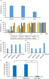
Hemolysis and pain induction by svAMPs. (A) Hemolytic activity of the crude venom of M. eupeus on mouse, lizard and bird erythrocytes. (B) Hemolysis of mouse erythrocytes by the svAMPs as a function of peptide concentration. Melittin was used as positive control. Hemolysis of lizard (C) and bird (D) erythrocytes by the peptides at 25 μM. Erythrocytes were suspended in PBS buffer and incubated with different concentrations of peptide for 30 min at 37°C. The absorbance of the supernatant was recorded at 570 nm. Controls for 0 and 100% hemolysis were determined by PBS buffer and 1% Triton X-100, respectively. Assays were repeated in triplicate and percentages of hemolysis are expressed as mean ± S.D. (E) Times of licking paw of mice induced by melittin and marmelittin, recorded between 5 and 30 min post-injection of peptides (n = 3). 0.9% NaCl was used as control. Scale bars represent means from three independent experiments; error bars, standard deviations (SDs). ***P < 0.001 (compared with the control without treatment by the peptides).
Figure 8
Figure 8
Dose-response curves resulting from injection of peptides into housefly adults. Data points are the mean ± SD of three experiments. LD50 of each svAMP was calculated from the dose-response curves. Melittin was used as control.
Figure 9
Figure 9
Sructural and functional features of MeuTXKβ1. (A) The sequence of MeuTxKβ1. Two distinct domains are highlighted in color and three disulfide bridges are also shown here. (B) Comparison of CD spectra between native and recombinant MeuTxKβ1 (Zhu et al., 2010). The recombinant product was a mixture of the full-length peptide and a truncated peptide with the N-terminal three residues (Gly-Phe-Arg) removed (Zhu et al., 2010). Peptide concentrations used here were 0.3 mg/ml. A minor negative band around 222 nm in the native peptide is indicated by an arrow. (C) Comparison of the anti-B. megaterium activity between MeuTXKβ1 and Meucin-18. (D) Killing kinetics of MeuTXKβ1. The S. aureus PRSA P1383 bacteria were treated with peptide solutions (specified in the figure, at 5x CL) for 5 to 60 min and survivors were plated. (E). Membrane permeation ability of MeuTXKβ1 on S. aureus PRSA P1383 at 5x CL. In (D,E), vancomycin was parallelly evaluated (5x CL for P1383 = 20 μg/ml) for comparison purpose. (F) Scanning electron microscopic observation of MeuTXKβ1-induced bacterial deformation. (G) Hemolysis of MeuTXKβ1 on mouse erythrocytes. Melittin was used as control. (H) The stability of MeuTXKβ1. Peptides were incubated in H2O or mouse serum for the indicated times and then added to bacterial plates containing S. aureus PRSA P1383. Diameters of inhibition zone were recorded after incubation at 37°C overnight. In (G,H), the data are presented as mean ± SD from three independent experiments. **P < 0.01; ***P < 0.001 (compared with the control without incubation in H2O or the serum).
Figure 10
Figure 10
The biological functions of svAMPs. These include bacterial killing, insect toxicity, hemolysis and pain induction, which are involved in predation and defense of scorpions against both predators and microbes. The crude venom was extracted from the M. eupeus telson by electrostimulation.

Similar articles

Cited by

References

    1. Almaaytah A., Albalas Q. (2014). Scorpion venom peptides with no disulfide bridges: a review. Peptides 51, 35–45. 10.1016/j.peptides.2013.10.021 - DOI - PubMed
    1. Arpornsuwan T., Buasakul B., Jaresitthikunchai J., Roytrakul S. (2014). Potent and rapid antigonococcal activity of the venom peptide BmKn2 and its derivatives against different Maldi biotype of multidrug-resistant Neisseria gonorrhoeae. Peptides 53, 315–320. 10.1016/j.peptides.2013.10.020 - DOI - PubMed
    1. Asthana N., Yadav S. P., Ghosh J. K. (2004). Dissection of antibacterial and toxic activity of melittin. A leucine zipper motif plays a crucial role in determining its hemolytic activity but not antibacterial activity. J. Biol. Chem. 279, 55042–55050. 10.1074/jbc.M408881200 - DOI - PubMed
    1. Barreiro L. B., Laval G., Quach H., Patin E., Quintana-Murci L. (2008). Natural selection has driven population differentiation in modern humans. Nat. Genet. 40, 340–345. 10.1038/ng.78 - DOI - PubMed
    1. Boland M. P., Separovic F. (2006). Membrane interactions of antimicrobial peptides from Australian tree frogs. Biochim. Biophys. Acta 1758, 1178–1183. 10.1016/j.bbamem.2006.02.010 - DOI - PubMed

LinkOut - more resources