. 2018 Mar 15:9:455.
doi: 10.3389/fmicb.2018.00455. eCollection 2018.

Evaluation of the Efficacy and Cross-Protective Immunity of Live-Attenuated Chimeric PCV1-2b Vaccine Against PCV2b and PCV2d Subtype Challenge in Pigs

Affiliations

Evaluation of the Efficacy and Cross-Protective Immunity of Live-Attenuated Chimeric PCV1-2b Vaccine Against PCV2b and PCV2d Subtype Challenge in Pigs

Changchao Huan et al. Front Microbiol. .

Abstract

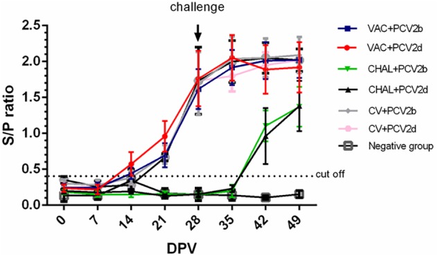
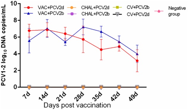
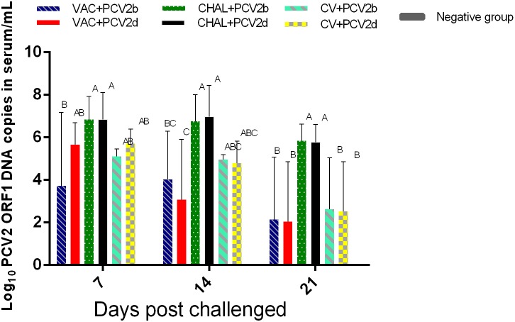
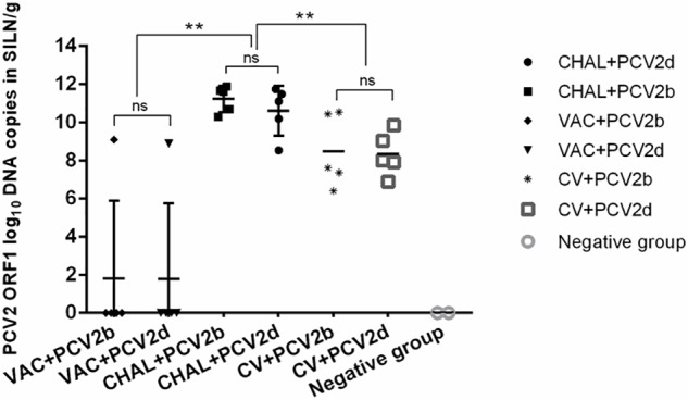
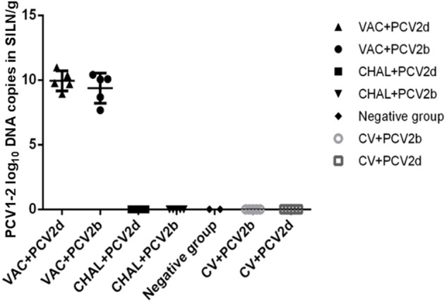
Porcine circovirus type 2 (PCV2) commercial vaccines are either inactivated PCV2 isolates or subunit vaccine based on the Cap protein of PCV2. Currently, no live-attenuated vaccines are yet available. Although the predominant circulating subtype worldwide is PCV2b, the emerging PCV2d subtype is also increasingly associated with PCV disease. In this study, piglets were inoculated with a live-attenuated chimeric PCV1-2b vaccine before challenged with PCV2b and PCV2d isolates. Thirty-two piglets were randomly divided into seven groups: negative (sham-vaccinated, sham-challenged), VAC+PCV2b (PCV1-2b vaccinated, PCV2b-challenged), VAC+PCV2d (PCV1-2b vaccinated, PCV2d-challenged), CHAL+PCV2b (sham-vaccinated, PCV2b-challenged), CHAL+PCV2d (sham-vaccinated, PCV2d-challenged), CV+PCV2b (commercial-vaccinated, PCV2b-challenged), and CV+PCV2d (commercial-vaccinated, PCV2d-challenged). The results showed that vaccinated challenged groups demonstrated high levels of anti-PCV2 antibody and reduced PCV2b and PCV2d loads both in serum and nasal swabs compared with the challenge-only groups. PCV2 DNA was detected in the superficial inguinal lymph nodes of only one pig in each of the VAC+PCV2b and VAC+PCV2d groups (group mean values, 101.81 and 101.77 genomic copies/g, respectively), which was significantly lower than those in CHAL+PCV2b and CHAL+PCV2d animals (group mean values, 1011.65 and 1010.60 genomic copies/g, respectively; P < 0.01). In addition, PCV2 DNA in each of the VAC+PCV2b and VAC+PCV2d groups was significantly lower than those in CV+PCV2b and CV+PCV2d animals (group mean values, 108.47 and 108.34 genomic copies/g, respectively; P < 0.01), indicating that the live-attenuated PCV1-2b vaccine was more effective than commercial vaccine. The live-attenuated PCV1-2b vaccine was effective in reducing PCV2b/PCV2d viremia, shedding, and tissue viral loads in vaccinated challenged pigs compared with challenge-only piglets, indicating that the PCV1-2b prototype vaccine is a good candidate for a live-attenuated vaccine against both PCV2b and PCV2d subtypes. And we first revealed that the live-attenuated PCV1-2b vaccine could protect piglets from challenge with either PCV2b or PCV2d equivalently.

Keywords: cross-protective immunity; live-attenuated chimeric porcine circovirus 1-2b; porcine circovirus 2b; porcine circovirus 2d; vaccine.


Figures

FIGURE 1
FIGURE 1
The sample-to-positive (S/P) ratio of each group at different days post vaccination (DPV) by PCV2 ELISA.
FIGURE 2
FIGURE 2
Group mean log10 of PCV1-2b genomic copies/mL in serum obtained from pigs at different days post vaccination (DPV).
FIGURE 3
FIGURE 3
Group mean log10 of PCV2 genomic copies/mL in serum obtained from pigs at different days post challenge (DPC). Different superscripts (A, B, C) indicate significant differences among groups (P < 0.05).
FIGURE 4
FIGURE 4
PCV2 DNA loads (copies/g) in SILN tissues collected on the day of necropsy (ns, P > 0.05; ∗∗P < 0.01).
FIGURE 5
FIGURE 5
PCV1-2b DNA loads (copies/g) in SILN tissues collected on the day of necropsy.
FIGURE 6
FIGURE 6
Histopathological lesions in experimental pigs. (A) No remarkable microscopic lesions were found in the lungs of the negative group pigs. (B) Slight interstitial pneumonia was found in the lung of one of five VAC+PCV2b groups pigs. (C) Slight lymphoplasmacytic and histiocytic bronchointerstitial pneumonia was present in the lungs of two of five VAC+PCV2d group pigs. (D) Severe lymphoplasmacytic and interstitial pneumonia was found in the lungs of four of five unvaccinated PCV2b-challenged pigs. (E) Severe interstitial pneumonia was observed in the lungs of four of five unvaccinated PCV2d-challenged pigs. (F) Moderate lymphoplasmacytic and interstitial pneumonia was observed in the lung two of five CV+PCV2b group pigs. (G) Moderate lymphoplasmacytic and interstitial pneumonia was observed in the lung two of five CV+PCV2d group pigs. (H) No remarkable microscopic lesions were found in the lymph nodes of pigs in the mock group. (I) No remarkable microscopic lesions were found in the lymph nodes of VAC+PCV2b group pigs. (J) Slight lymphocyte deletion was observed in the lymph node follicles of two of five VAC+PCV2d group pigs. (K) Severe macrophage accumulation was found in lymph node follicles of all five unvaccinated PCV2b-challenged pigs. (L) Severe lymphoid depletion (LD) was observed in lymph node follicles of four of five unvaccinated PCV2d-challenged pigs. (M) Severe eosinophil infiltration in the lymph nodes in one pig of CV+PCV2b group. (N) LD in lymph nodes follicles in one pig of CV+PCV2d group.
FIGURE 7
FIGURE 7
Immunohistochemistry detection of PCV2 antigen in SILN. (A) PCV2 antigen was not detected in lymph nodes of mock pigs. (B) Sparse staining for PCV2 antigen was detectable in the lymph nodes of VAC+PCV2b pigs. (C) Sparse staining for PCV2 antigen was detectable in lymph nodes of VAC+PCV2d pigs. (D) Strong staining for PCV2 antigen was evident in lymph nodes of CHAL+PCV2b pigs. (E) Strong staining for PCV2 antigen was evident in lymph nodes of CHAL+PCV2d pigs. (F) Sparse staining for PCV2 antigen was detectable in the lymph nodes of CV+PCV2b pigs. (G) Sparse staining for PCV2 antigen was detectable in lymph nodes of CV+PCV2d pigs.
FIGURE 8
FIGURE 8
Fluorescence in situ hybridization detection of PCV2b and PCV2d DNA in SILN. (A) PCV2b DNA was not detected in lymph nodes of mock pigs. (B) PCV2d DNA was not detected in lymph nodes of mock pigs. (C) Strong staining for PCV2b DNA was evident in lymph nodes of CHAL+PCV2b pigs. (D) Sparse staining for PCV2b DNA was detectable in the lymph nodes of CV+PCV2b pigs. (E) Sparse staining for PCV2b DNA was detectable in the lymph nodes of VAC+PCV2b pigs. (F) Strong staining for PCV2d DNA was evident in lymph nodes of CHAL+PCV2d pigs. (G) Sparse staining for PCV2d DNA was detectable in lymph nodes of CV+PCV2d pigs. (H) Sparse staining for PCV2d DNA was detectable in lymph nodes of VAC+PCV2d pigs.

Similar articles

Cited by

References

    1. Aida J., Izumiyama-Shimomura N., Nakamura K., Ishii A., Ishikawa N., Honma N., et al. (2007). Telomere length variations in 6 mucosal cell types of gastric tissue observed using a novel quantitative fluorescence in situ hybridization method. Hum. Pathol. 38 1192–1200. 10.1016/j.humpath.2006.11.023 - DOI - PubMed
    1. Allan G. M., Ellis J. A. (2000). Porcine circoviruses: a review. J. Vet. Diagn. Invest. 12 3–14. 10.1177/104063870001200102 - DOI - PubMed
    1. Beach N. M., Ramamoorthy S., Opriessnig T., Wu S. Q., Meng X. J. (2010). Novel chimeric porcine circovirus (PCV) with the capsid gene of the emerging PCV2b subtype cloned in the genomic backbone of the non-pathogenic PCV1 is attenuated in vivo and induces protective and cross-protective immunity against PCV2b and PCV2a subtypes in pigs. Vaccine 29 221–232. 10.1016/j.vaccine.2010.10.050 - DOI - PubMed
    1. Davies B., Wang X., Dvorak C. M. T., Marthaler D., Murtaugh M. P. (2016). Diagnostic phylogenetics reveals a new Porcine circovirus 2 cluster. Virus Res. 217 32–37. 10.1016/j.virusres.2016.02.010 - DOI - PubMed
    1. Dupont K., Nielsen E. O., Baekbo P., Larsen L. E. (2008). Genomic analysis of PCV2 isolates from Danish archives and a current PMWS case-control study supports a shift in genotypes with time. Vet. Microbiol. 128 56–64. 10.1016/j.vetmic.2007.09.016 - DOI - PubMed

LinkOut - more resources