. 2018 Jan 21:2018:1023025.
doi: 10.1155/2018/1023025. eCollection 2018.

Thioredoxin-1 Protects Bone Marrow-Derived Mesenchymal Stromal Cells from Hyperoxia-Induced Injury In Vitro

Affiliations

Thioredoxin-1 Protects Bone Marrow-Derived Mesenchymal Stromal Cells from Hyperoxia-Induced Injury In Vitro

Lei Zhang et al. Oxid Med Cell Longev. .

Abstract

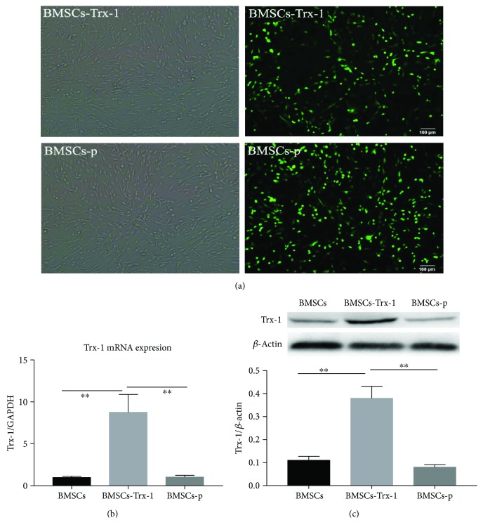
Background: The poor survival rate of mesenchymal stromal cells (MSC) transplanted into recipient lungs greatly limits their therapeutic efficacy for diseases like bronchopulmonary dysplasia (BPD). The aim of this study is to evaluate the effect of thioredoxin-1 (Trx-1) overexpression on improving the potential for bone marrow-derived mesenchymal stromal cells (BMSCs) to confer resistance against hyperoxia-induced cell injury.

Methods: 80% O2 was used to imitate the microenvironment surrounding-transplanted cells in the hyperoxia-induced lung injury in vitro. BMSC proliferation and apoptotic rates and the levels of reactive oxygen species (ROS) were measured. The effects of Trx-1 overexpression on the level of antioxidants and growth factors were investigated. We also investigated the activation of apoptosis-regulating kinase-1 (ASK1) and p38 mitogen-activated protein kinases (MAPK).

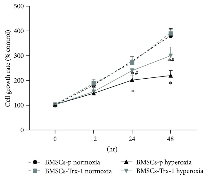
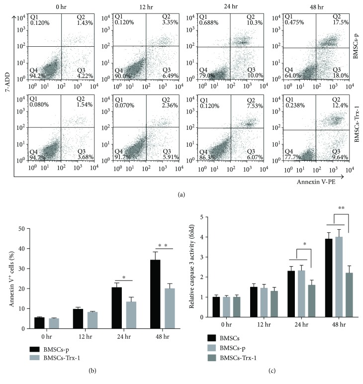
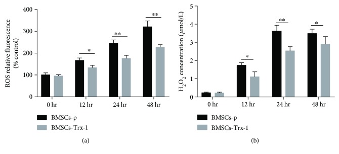
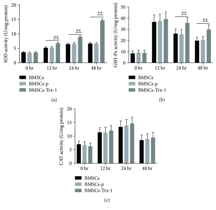
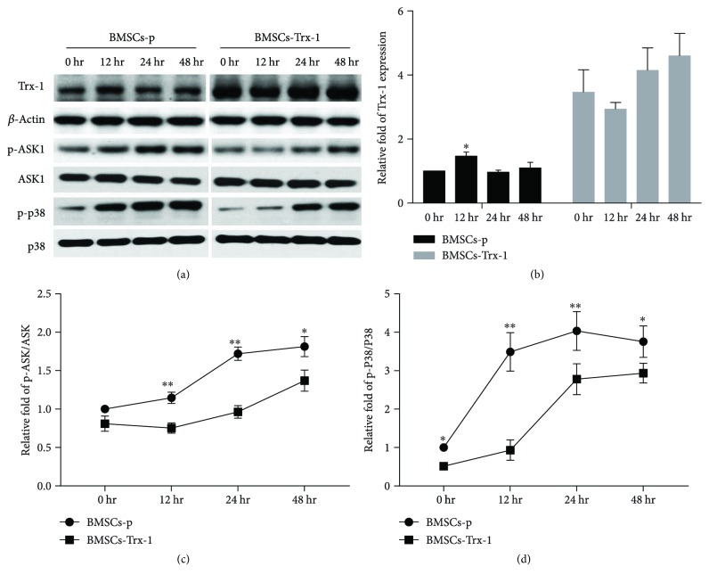
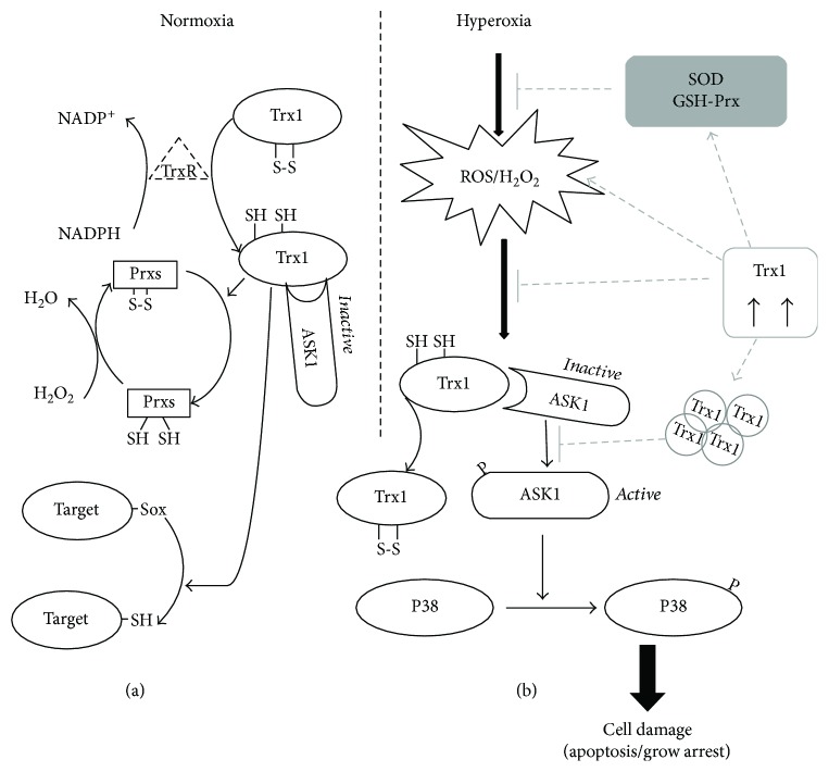
Result: Trx-1 overexpression significantly reduced hyperoxia-induced BMSC apoptosis and increased cell proliferation. We demonstrated that Trx-1 overexpression upregulated the levels of superoxide dismutase and glutathione peroxidase as well as downregulated the production of ROS. Furthermore, we illustrated that Trx-1 protected BMSCs against hyperoxic injury via decreasing the ASK1/P38 MAPK activation rate.

Conclusion: These results demonstrate that Trx-1 overexpression improved the ability of BMSCs to counteract hyperoxia-induced injury, thus increasing their potential to treat hyperoxia-induced lung diseases such as BPD.


Figures

Figure 1
Figure 1
Characterization of rat bone marrow-derived mesenchymal stromal cells (BMSCs). (a) The plastic-adherent cells demonstrated a homogeneous fibroblast-like and spindle-shaped morphology. Original magnification, ×100. (b) Adipogenic differentiation of BMSCs stained with oil red O. Original magnification, ×200. (c) Osteogenic differentiation of BM-MSCs stained with alizarin red. Original magnification, ×400. (d) FACS analysis demonstrated expression of markers attributed to BMSCs. The cells were devoid of hematopoietic cells as indicated by the lack of CD45 and CD34. The MSC-specific markers, CD29, CD44, CD73, CD105, and CD90 were strongly expressed on the cells.
Figure 2
Figure 2
Stable overexpression of Trx-1 in BMSCs. (a) Intensive green fluorescence was observed by fluorescence microscopy (×100). (b) The mRNA levels of Trx-1 in BMSCs, BMSCs-Trx-1, and BMSCs-p. (c) Detection of Trx-1 protein expression by Western blot analysis. ∗∗ P < 0.01 compared to control. BMSCs: intact BMSCs; BMSCs-p: empty lentivirus-engineered BMSCs; BMSCs-Trx-1: Trx-1-engineered BMSCs.
Figure 3
Figure 3
Overexpression of Trx-1 promoted proliferation of BMSCs under hyperoxic conditions. Cells with or without Trx-1 overexpression were exposed to hyperoxia for the indicated time. Cell proliferation was estimated using a CCK-8 kit. Hyperoxia treatment inhibited BMSC proliferation. However, overexpression of Trx-1 increased cell growth rate under hyperoxic conditions compared to BMSCs-p. Growth curve was generated by reading the absorbance value at 450 nm. The value was computed as percent of 0 hour. The results were expressed as mean ± SD of the results of three independent experiments, each with triplicates. P < 0.05 or 0.01 compared to normoxia control, # P < 0.05 or 0.01 compared to BMSCs-p under hyperoxia conditions. BMSCs-p: empty lentivirus-engineered BMSCs; BMSCs-Trx-1: Trx-1-engineered BMSCs.
Figure 4
Figure 4
Effect of Trx-1 on cell apoptosis in BMSCs. Cells were exposed to hyperoxia for 0, 12, 24, and 48 hours and were stained with annexin V-PE/7-ADD before flow cytometry analysis. (a) Dot plots of flow cytometry analysis. Intensity of 7-ADD staining (y-axis) was plotted versus annexin V intensity (x-axis). Numbers indicate percent in each region. (b) The graph shows the percentage of apoptosis as defined by annexin V+. The results are representative of 3 independent experiments. (c) Caspase 3 activity. Caspase 3 activity was measured by the caspase 3 activity kit. Bar graphs represent the relative expression of caspase 3 activity calculated from each group. The results are representative of 3 independent experiments. P < 0.05, ∗∗ P < 0.01 compared with the BMSCs-p group or BMSCs. BMSCs: intact BMSCs; BMSCs-p: empty lentivirus-engineered BMSCs; BMSCs-Trx-1: Trx-1-engineered BMSCs.
Figure 5
Figure 5
Effects of Trx-1 on intracellular ROS levels in BMSCs. (a) Intracellular ROS production was measured with CellROX deep red reagent, which can detect total ROS and was not the target particular species. The relative fluorescence intensity was expressed as % compared to control cells (BMSCs-p at 0 hr). (b) The level of intracellular H2O2 was measured using hydrogen peroxide assay kit. Experiments were repeated three times. P < 0.05, ∗∗ P < 0.01 versus the corresponding group. BMSCs-p: empty lentivirus-engineered BMSCs; BMSCs-Trx-1: Trx-1-engineered BMSCs.
Figure 6
Figure 6
Effects of Trx-1 overexpression on antioxidant enzyme activities in BMSCs under hyperoxic conditions. (a) Superoxide dismutase (SOD) activities were measured using the SOD assay kit. (b) Glutathione peroxidase (GSH-Px) activities were measured using the glutathione peroxidase assay kit. (c) Catalase (CAT) activities were measured using the CAT assay kit. Data are representative of duplicate samples from five experiments. ∗∗ P < 0.01. BMSCs: intact BMSCs; BMSCs-p: empty lentivirus-engineered BMSCs; BMSCs-Trx-1: Trx-1-engineered BMSCs.
Figure 7
Figure 7
Western blot results. Trx-1, phospho-ASK1, total ASK1, phospho-p38, and total p38 expressions were detected by Western blotting. (a) Representative Western blot bands. (b) Trx-1 densitometric analysis. (c) p-ASK/ASK densitometric analysis. (d) p-38/P38 densitometric analysis. Data are representative of three independent experiments. BMSCs-p: empty lentivirus-engineered BMSCs; BMSCs-Trx-1: Trx-1-engineered BMSCs. P < 0.05; ∗∗ P < 0.01 versus the corresponding group.
Figure 8
Figure 8
A schematic model of the regulation of the ASK1/P38 signal pathway by Trx-1. (a) The Trx-1 system contains NADPH, TrxR-1, and Trx-1. The oxidized Trx-1 (inactive form) is transformed to the active and reduced form of Trx-1 by receiving electrons from NADPH coenzyme in the presence of TrxR-1. Prxs reduce H2O2 to H2O using electrons from the active Trx-1. The active Trx-1 also regulates redox signals by reducing many other target proteins with disulfide bonds. ASK1 constantly forms an inactive complex with reduced Trx-1 under normoxic conditions. (b) Exposure of BMSCs to hyperoxia leads to elevated ROS and H2O2 production, which leads to oxidative stress. However, oxidized Trx-1 is dissociated from ASK1 in response to oxidative stress and subsequent activation of ASK1. Activated ASK1 in turn activates the p38 pathway and induces various cellular responses, including cell apoptosis and differentiation inhibition. Trx-1 overexpression promoted BMSC survival under hyperoxic conditions through elevation of antioxidant activities, reduction of ROS and H2O2 generation, and subsequent inhibition of the ASK1/P38 signaling pathway.

Similar articles

Cited by

References

    1. Madurga A., Mizikova I., Ruiz-Camp J., Morty R. E. Recent advances in late lung development and the pathogenesis of bronchopulmonary dysplasia. American Journal of Physiology Lung Cellular and Molecular Physiology. 2013;305(12):L893–L905. doi: 10.1152/ajplung.00267.2013. - DOI - PubMed
    1. Frank L., Massaro D. Oxygen toxicity. The American Journal of Medicine. 1980;69(1):117–126. doi: 10.1016/0002-9343(80)90509-4. - DOI - PubMed
    1. Thomson L., Paton J. Oxygen toxicity. Paediatric Respiratory Reviews. 2014;15(2):120–123. doi: 10.1016/j.prrv.2014.03.003. - DOI - PubMed
    1. Boeckel D. G., Shinkai R. S. A., Grossi M. L., Teixeira E. R. Cell culture–based tissue engineering as an alternative to bone grafts in implant dentistry: a literature review. Journal of Oral Implantology. 2012;38(S1):538–545. doi: 10.1563/AAID-JOI-D-11-00197. - DOI - PubMed
    1. Wang J., Xu L., Chen Q., Zhang Y., Hu Y., Yan L. Bone mesenchymal stem cells overexpressing FGF4 contribute to liver regeneration in an animal model of liver cirrhosis. International Journal of Clinical and Experimental Medicine. 2015;8(8):12774–12782. - PMC - PubMed

LinkOut - more resources