Review
. 2018 Feb 11:2018:3075293.
doi: 10.1155/2018/3075293. eCollection 2018.

Emerging Players at the Intersection of Chondrocyte Loss of Maturational Arrest, Oxidative Stress, Senescence and Low-Grade Inflammation in Osteoarthritis

Affiliations
Review

Emerging Players at the Intersection of Chondrocyte Loss of Maturational Arrest, Oxidative Stress, Senescence and Low-Grade Inflammation in Osteoarthritis

Manuela Minguzzi et al. Oxid Med Cell Longev. .

Abstract

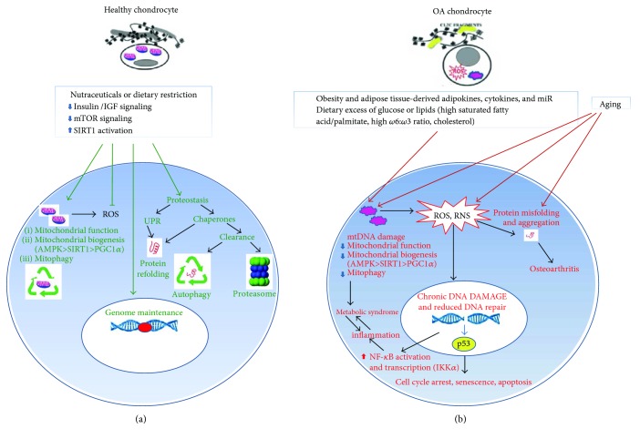
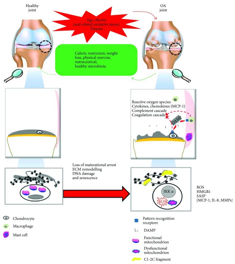
The prevalence of Osteoarthritis (OA) is increasing because of the progressive aging and unhealthy lifestyle. These risk factors trigger OA by removing constraints that keep the tightly regulated low turnover of the extracellular matrix (ECM) of articular cartilage, the correct chondrocyte phenotype, and the functionality of major homeostatic mechanisms, such as mitophagy, that allows for the clearance of dysfunctional mitochondria, preventing increased production of reactive oxygen species, oxidative stress, and senescence. After OA onset, the presence of ECM degradation products is perceived as a "danger" signal by the chondrocytes and the synovial macrophages that release alarmins with autocrine/paracrine effects on the same cells. Alarmins trigger innate immunity in the joint, with important systemic crosstalks that explain the beneficial effects of dietary interventions and improved lifestyle. Alarmins also boost low-grade inflammation: the release of inflammatory molecules and chemokines sustained by continuous triggering of NF-κB within an altered cellular setting that allows its higher transcriptional activity. Chemokines exert pleiotropic functions in OA, including the recruitment of inflammatory cells and the induction of ECM remodeling. Some chemokines have been successfully targeted to attenuate structural damage or pain in OA animal models. This represents a promising strategy for the future management of human OA.


Figures

Figure 1
Figure 1
Failure of major homeostatic mechanisms contributes to OA pathogenesis. Failure of major homeostatic mechanisms (proteostasis and DNA damage repair networks and mitochondrial respiratory metabolism) in articular chondrocytes is caused by aging or conditions that accelerate tissue aging, such as the obesity-associated systemic low-grade inflammation and/or dietary factors (dyslipidemia and/or hyperglycemia) that impact on mitochondrial function. (a) Homeostasis is tightly controlled by balance between the insulin/IGF_mTOR signaling and SIRT1. In conditions of homeostasis normal mitochondrial function, biogenesis and autophagy (mitophagy) guarantee that the level of ROS is kept to the minimum required for intracellular signaling. Both genomic and mtDNA are preserved from oxidative damage. Proteostasis is guaranteed by the correct functioning of the unfolded protein response and clearance via autophagy or the ubiquitin-proteasome system. (b) Deranged metabolic factors together with aging contribute to mitochondrial dysfunction, accumulation of ROS and RNS that increase the level of protein misfolding and aggregation, and impact on the integrity of both mitochondrial and genomic DNA. Accumulation of DNA damage cannot be efficiently corrected because mitochondrial dysfunction leads to failure of the energy supply required by the DNA damage response. Persistent DNA damage is responsible for chronic NF-κB activation, and inflammation, leading to the “metabolic syndrome.” A positive feedback loop between metabolic syndrome and inflammation is even worsened by excessive ROS and RNS produced by the dysfunctional mitochondria. Persistent DNA damage is also responsible for p53 activation, with functional consequences for the cells that include cell cycle arrest, senescence, or apoptosis according to an increasing degree.
Figure 2
Figure 2
(a) Sources of ROS in the chondrocyte. The major sources of ROS are the mitochondria, during the OXPHOS. In addition, ROS can be generated at the level of the endoplasmic reticulum (ER), in condition of ER stress. An additional source is represented by the peroxisome, during the β-oxidation of long chain-fatty acids (LCFA), particularly under conditions of excess LCFA load. Various enzymes, with different cell location may also generate ROS. (b) Production of ROS and RNS involved in OA pathogenesis, and major detoxifying enzymes. The first generated ROS is the superoxide (O2) that is either rapidly transformed into hydrogen peroxide (H2O2) by the enzymes of the superoxide dismutase (SOD) family or can act as a precursor for peroxynitrite (ONOO) formation when joined to nitric oxide (NO), produced by the enzymes of the nitric oxide synthase (NOS) family. Because of unpaired electrons, free radicals are very reactive and can damage cell components. In chondrocytes, iron Fe2+ and H2O2 release hydroxyl radicals (OH) that react with unsaturated fatty acids of membrane lipids, thus forming lipid radicals (RO, ROO). ROS are neutralized by scavenging systems, namely, SOD, catalase, glutathione peroxidase, glutathione reductase, and reduced glutathione.
Figure 3
Figure 3
Macro to micro views of changes occurring in articular cartilage during OA onset and development. Left images indicate healthy joint, joint tissues, and chondrocytes. Resting chondrocytes present functional mitochondria and are surrounded by intact ECM. As a consequence of different risk factors, profound changes occur in all the joint tissues. Right images indicate OA joint, crosstalk among OA joint tissues, and hypertrophic chondrocytes. The latter present dysfunctional mitochondria, increased ROS production, and increased IKKα expression and are surrounded by degraded ECM. ECM degradation products act as damage associated molecular patterns (DAMPs or alarmins) recognized by the pattern recognition receptors (PRR) expressed by the cells. Alarmins are able to trigger both synovial macrophages (via TLR2 and TLR4) and chondrocytes (via TLR2 and TLR4, and RAGE), thus sustaining synovial inflammation and cartilage degradation. DAMPs also include HMGB1 that is highly expressed by both OA synoviocytes and hypertrophic chondrocytes and is able to enhance NF-κB activation and release of chemokines (particularly of MCP-1, highly active in the recruitment of monocytes that are also present in the synovial fluid) and cytokines, as well as of matrix metalloproteinases (MMPs) responsible for ECM remodeling and loss of maturational arrest. Increased oxidative stress is both cause and consequence of mitochondrial dysfunction. Correction of major risk factors and changes in diet and lifestyle can significantly reduce disease progression.

Similar articles

Cited by

References

    1. Wallace I. J., Worthington S., Felson D. T., et al. Knee osteoarthritis has doubled in prevalence since the mid-20th century. Proceedings of the National Academy of Sciences of the United States of America. 2017;114(35):9332–9336. doi: 10.1073/pnas.1703856114. - DOI - PMC - PubMed
    1. Miyaki S., Asahara H. Macro view of microRNA function in osteoarthritis. Nature Reviews Rheumatology. 2012;8(9):543–552. doi: 10.1038/nrrheum.2012.128. - DOI - PMC - PubMed
    1. Haigis M. C., Yankner B. A. The aging stress response. Molecular Cell. 2010;40(2):333–344. doi: 10.1016/j.molcel.2010.10.002. - DOI - PMC - PubMed
    1. Franceschi C., Garagnani P., Vitale G., Capri M., Salvioli S. Inflammaging and ‘Garb-aging’. Trends in Endocrinology and Metabolism. 2017;28(3):199–212. doi: 10.1016/j.tem.2016.09.005. - DOI - PubMed
    1. Lourido L., Ayoglu B., Fernandez-Tajes J., et al. Discovery of circulating proteins associated to knee radiographic osteoarthritis. Scientific Reports. 2017;7(1):p. 137. doi: 10.1038/s41598-017-00195-8. - DOI - PMC - PubMed

LinkOut - more resources