

Three-dimensional computed tomography reconstruction for operative planning in robotic segmentectomy: a pilot study
- PMID: 29600049
- PMCID: PMC5863115
- DOI: 10.21037/jtd.2017.11.144
Three-dimensional computed tomography reconstruction for operative planning in robotic segmentectomy: a pilot study
Abstract
Background: The objective of our pilot study was to assess if three-dimensional (3D) reconstruction performed by Visible Patient™ could be helpful for the operative planning, efficiency and safety of robot-assisted segmentectomy.
Methods: Between 2014 and 2015, 3D reconstructions were provided by the Visible Patient™ online service and used for the operative planning of robotic segmentectomy. To obtain 3D reconstruction, the surgeon uploaded the anonymized computed tomography (CT) image of the patient to the secured Visible Patient™ server and then downloaded the model after completion.
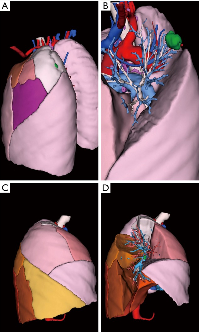
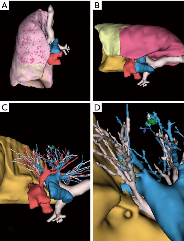
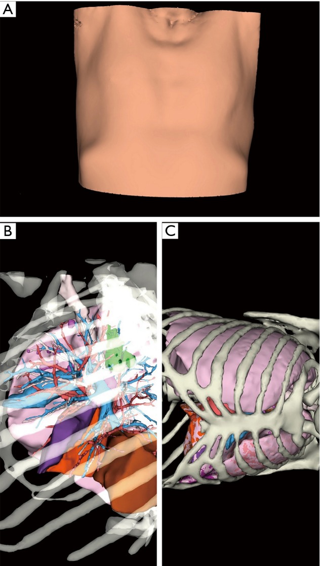
Results: Nine segmentectomies were performed between 2014 and 2015 using a pre-operative 3D model. All 3D reconstructions met our expectations: anatomical accuracy (bronchi, arteries, veins, tumor, and the thoracic wall with intercostal spaces), accurate delimitation of each segment in the lobe of interest, margin resection, free space rotation, portability (smartphone, tablet) and time saving technique.
Conclusions: We have shown that operative planning by 3D CT using Visible Patient™ reconstruction is useful in our practice of robot-assisted segmentectomy. The main disadvantage is the high cost. Its impact on reducing complications and improving surgical efficiency is the object of an ongoing study.
Keywords: Robotic surgery; ground-glass nodules; lung cancer; segmentectomy; three-dimensional computed tomography (3D CT) reconstruction.
Conflict of interest statement
Conflicts of Interest: JM Baste discloses fees for lecturing and proctoring for Intuitive Surgical. This work was presented at the French Thoracic and Cardio-Vascular Society autumn 2017 meeting.
Figures

References
-
- Detterbeck FC, Lewis SZ, Diekemper R, et al. Executive summary: Diagnosis and management of lung cancer, 3rd ed: american college of chest physicians evidence-based clinical practice guidelines. Chest 2013;143:7S–37S. - PubMed
-
- Linden D, Linden K, Oparka J. In patients with resectable non-small-cell lung cancer, is video-assisted thoracoscopic segmentectomy a suitable alternative to thoracotomy and segmentectomy in terms of morbidity and equivalence of resection? Interact Cardiovasc Thorac Surg 2014;19:107-10. 10.1093/icvts/ivu080 - DOI - PubMed
LinkOut - more resources
Full Text Sources
Other Literature Sources
Miscellaneous