

Scleral ultrastructure and biomechanical changes in rabbits after negative lens application
- PMID: 29600166
- PMCID: PMC5861222
- DOI: 10.18240/ijo.2018.03.02
Scleral ultrastructure and biomechanical changes in rabbits after negative lens application
Abstract
Aim: To address the microstructure and biomechanical changes of the sclera of rabbits after negative lens application by spectacle frame apparatus.
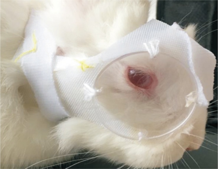
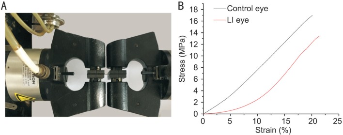
Methods: Five New Zealand rabbits of seven weeks post-natal were treated with -8 D lens monocularly over the course of two weeks. Refractive errors and axial length (AXL) were measured at the 1st, 7th and 14th days of the induction period. Ultrastructure of sclera was determined with electron microscopy. Biomechanical properties were tested by an Instron 5565 universal testing machine.
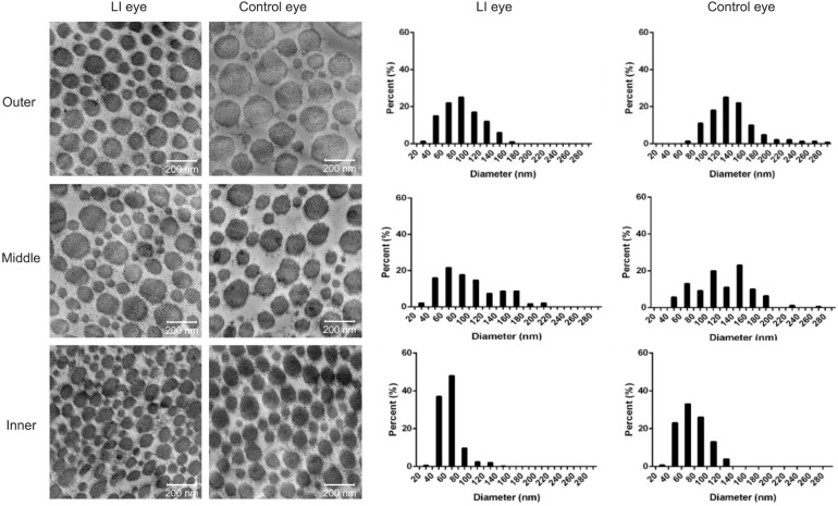
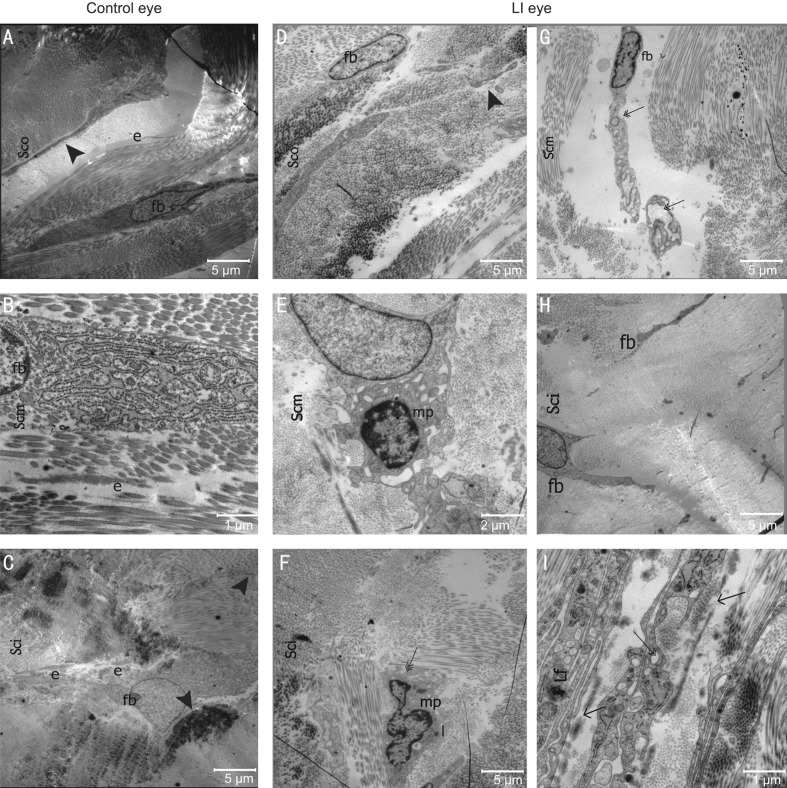
Results: Lens-induced (LI) eyes elongated more rapidly compared with fellow eyes with AXL values of 15.56±0.14 and 15.21±0.14 mm (P<0.01). Fibril diameter was significantly smaller in the LI eyes compared with control ones in the inner, middle, and outer layers (inner layer, 63.533 vs 76.467 nm; middle layer, 92.647 vs 123.984 nm; outer layer, 86.999 vs 134.257 nm, P<0.01, respectively). In comparison with control eyes, macrophage-like cells that engulfed fibroblasts, dilated endoplasmic reticulum, and vacuoles in fibroblasts were observed in the inner and middle stroma in the LI eyes. Ultimate stress and Young's modulus were lower in the LI eyes compared with those in the control eyes.
Conclusion: Negative lens application alters eye growth, and results in axial elongation with changes in scleral ultrastructural and mechanical properties.
Keywords: biomechanics; negative lens; rabbit; sclera; ultrastructure.
Figures

References
-
- Rada JA, Shelton S, Norton TT. The sclera and myopia. Exp Eye Res. 2006;82(2):185–200. - PubMed
-
- Lin Z, Chen X, Ge J, Cui D, Wu J, Tang F, Tan J, Zhong X, Gao Q. Effects of direct intravitreal dopamine injection on sclera and retina in form-deprived myopic rabbits. J Ocul Pharmacol Ther. 2008;24(6):543–550. - PubMed
-
- McBrien NA, Jobling AI, Gentle A. Biomechanics of the sclera in myopia: extracellular and cellular factors. Optom Vis Sci. 2009;86(1):23–30. - PubMed
LinkOut - more resources
Full Text Sources
Other Literature Sources
Research Materials
Miscellaneous