. 2018 Feb;3(1):97-109.
doi: 10.1016/j.jacbts.2017.09.005.

Diffusion Tensor Cardiac Magnetic Resonance Reveals Exosomes From Cardiosphere-Derived Cells Preserve Myocardial Fiber Architecture After Myocardial Infarction

Affiliations

Diffusion Tensor Cardiac Magnetic Resonance Reveals Exosomes From Cardiosphere-Derived Cells Preserve Myocardial Fiber Architecture After Myocardial Infarction

Christopher T Nguyen et al. JACC Basic Transl Sci. 2018 Feb.

Abstract

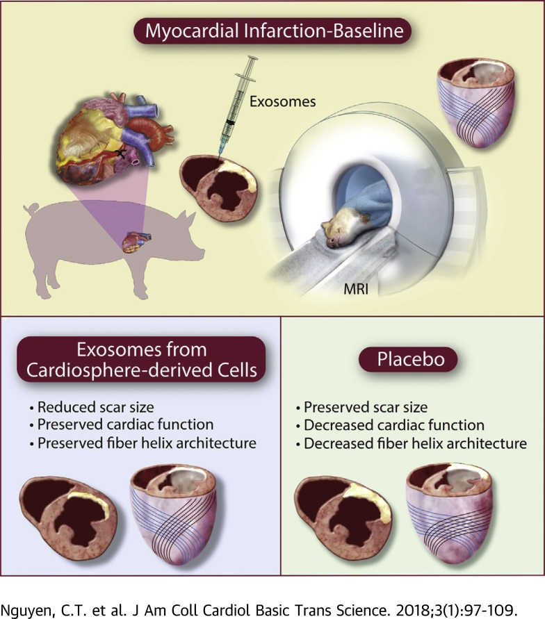
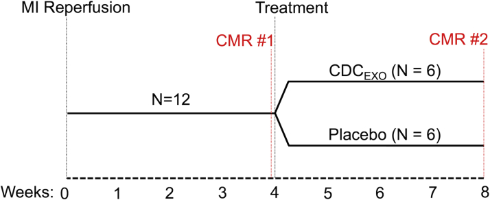
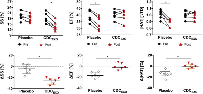
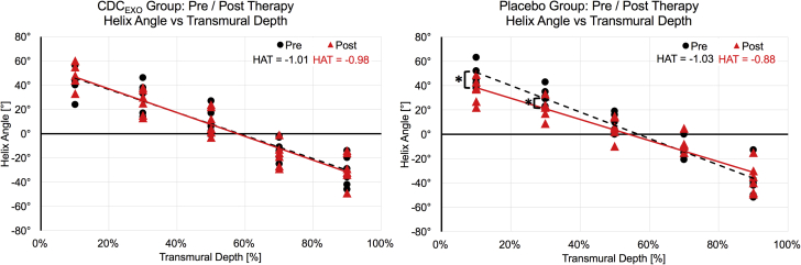
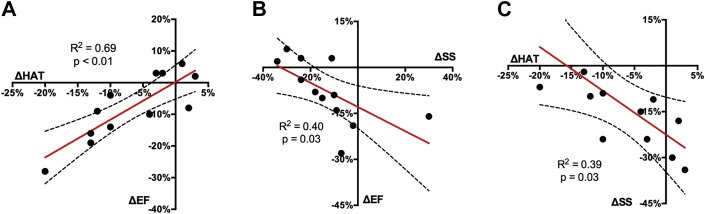
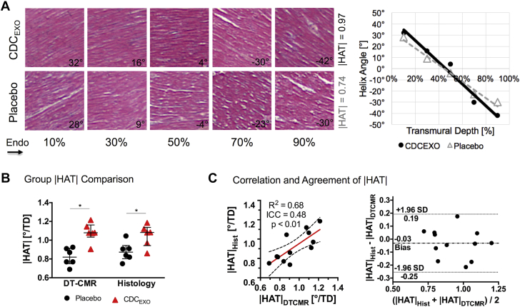
The object of the study was to reveal the fiber microstructural response with diffusion tensor cardiac magnetic resonance after intramyocardial exosomes secreted by cardiosphere-derived cells (CDCEXO) in chronic porcine myocardial infarction. Porcine with myocardial infarction underwent intramyocardial delivery of human CDCEXO and placebo in a randomized placebo-controlled study. Four weeks after injection, viability improved in the CDCEXO group, whereas myocardial fiber architecture and cardiac function were preserved. In the placebo group, fiber architecture and cardiac function declined. Myocardial regeneration by CDCEXO is not tumor-like; instead, details of tissue architecture are faithfully preserved, which may foster physiological excitation and contraction.

Keywords: diffusion tensor MRI; exosomes; fiber architecture; myocardial infarction; regeneration.


Figures

None
Graphical abstract
Figure 1
Figure 1
Study Design of the Randomized Placebo Controlled Study All subjects undergo MI reperfusion and after 4 weeks of recovery CMR is performed to establish baseline. After CMR scan 1, one-half the group is randomly chosen to be injected with CDCEXO and the other one-half with a placebo. Both groups undergo a second CMR scan after 4 weeks post-injection. CDCEXO = exosomes from cardiosphere-derived cells, CMR = cardiac magnetic resonance; MI = myocardial infarction.
Figure 2
Figure 2
Cartoon Representations of the Myocardial Fiber Architecture (A) Normal and (B) infarcted left ventricle. α characterizes the inclination of the estimated myocardial fiber orientation of each voxel in relation to the short axis plane (defined by r and t) and tangent plane (defined by t and u). For normal myocardial fiber architecture, the helix angle smoothly changes from the endocardium (red) to mid-myocardium (green) to epicardium (blue). This transmural change is roughly linear and can be characterized by fitting a slope through α plotted against transmural depth yielding the HAT. For infarcted myocardium, the myocardial fiber architecture in the remote region exhibits less right-handed fibers in the endocardium (α > 0) with a “flatter” overall HAT. (C) Representative image quality of the DW images from a single placebo subject before induced MI, baseline MI pre-injection, and post-injection. The DW images were used to reconstruct the diffusion tensor and subsequently used to derive HA. The white arrow points to the center of the scar. α = helix angle; DW = diffusion-weighted; HA = helix angle; HAT = helix angle transmurality.
Figure 3
Figure 3
Representative Images of LGE, CINE, HA Map, and |ΔHAT| p Value Wheel for Pre- and Post-CDCEXO and Placebo Groups For the CDCEXO subject, the red arrows point to a region of scar (anteroseptal) reduction in the LGE and a local restoration of the |HAT| with the endocardial fibers significantly (p < 0.05) changing from circumferential (HA ∼ 0) to right-handed helical (HA > 0) orientation (|HATpre| = 0.74°/%TD, |HATpost| = 0.95°/%TD). CINE revealed preserved LVEF. For the majority of the remote myocardium (anterolateral, inferolateral, inferior, and inferoseptal walls), |HAT| remained impaired with no significant change. For the placebo subject, LGE yielded no significant changes in scar size. CINE exhibited a reduction in LVEF. The |HAT| (white arrows) was significantly reduced in the inferoseptal wall with epicardial fibers changing from left-handed (HA < 0) to circumferential (HA ∼ 0) orientation (|HATpre| = 1.04°/%TD, |HATpost| = 0.78°/%TD). The |HAT| was also significantly reduced in the inferior wall with endocardial fibers changing from right-handed helical (HA > 0) to circumferential (HA ∼0) orientation (|HATpre| = 0.92°/%TD, |HATpost| = 0.69°/%TD). Anterolateral and inferolateral walls remained impaired with no significant change. CINE = cinematic magnetic resonance imaging; |HAT| = absolute helix angle transmurality; LGE = late gadolinium enhancement, TD = transmural depth; other abbreviation as in Figure 1.
Figure 4
Figure 4
Global Line Plots and Normalized Changes of SS, |HAT|, and EF for Both CDCEXO and Placebo Groups Global line plots (top) and normalized changes (bottom) demonstrate baseline values for SS, |HAT|, and EF were comparable between both groups. The CDCEXO group exhibited significant (p < 0.05) reduction in SS but no significant changes in |HAT| and EF. The placebo group revealed significantly reduced |HAT| and EF but no change in SS. EF = ejection fraction; SS = scar size; other abbreviations as in Figures 1 and 3.
Figure 5
Figure 5
HA Versus TD Plots for Pre/Post Scans for the CDCEXO and Placebo Groups The calculated HAT (slope of the HA vs. TD line plot) did not significantly change in the treated group (−1.01°/%TD to −0.98°/%TD). In contrast, the placebo group exhibited a significant (p = 0.03) reduction in HAT (−1.03°/%TD to −0.88°/%TD) between the pre/post scans indicating an overall loss in helix transmurality. Furthermore, HA was significantly (p = 0.03 and p = 0.04, respectively) different at 10% and 30% TD between pre/post scans, with 90% TD trending toward significance (p = 0.08). Abbreviation as in Figures 1 and 3.
Figure 6
Figure 6
Univariate Linear Regression Plots (A) ΔEF vs ΔHAT, (B) ΔEF vs ΔSS, and (C) ΔSS vs ΔHAT. ΔEF significantly (p < 0.01) and positively correlated (R2 = 0.69) with ΔHAT, but significantly (p = 0.03) and negatively correlated (R2 = 0.40) with ΔSS. ΔSS was significantly (p = 0.03) correlated (R2 = 0.39) with ΔHAT. ΔEF = change in left ventricular ejection fraction; ΔHAT = change in helix angle transmurality; ΔSS = change in scar size.
Figure 7
Figure 7
Histological-Derived |HAT| (A) Single representative case, (B) group comparison, and (C) correlation and agreement with DT-CMR–derived |HAT| values. The stained hematoxylin and eosin histological sections clearly show the orientation of the myocardial fibers at varying transmural depths, which can be quantified to yield a helix angle versus transmural depth plot and HAT values. Both DT-CMR and histology were concordant in exhibiting the same trend of a significant decreased |HAT| in the placebo group compared with the CDCEXO-treated group. Furthermore, substantial correlation and agreement were found between histology and DT-CMR–derived |HAT| demonstrated in the correlation and Bland-Altman plot. DT-CMR = diffusion-tensor cardiac magnetic resonance. Abbreviation as in Figures 1 and 3.
Figure 8
Figure 8
ROC Curves of ΔSS, ΔHAT, and Combined ΔSS and ΔHAT to Predict ΔEF ΔHAT and ΔSS had comparable predictive values that did not significantly differ (AUC = 0.66 and 0.68, respectively). Combining ΔSS and ΔHAT with the best fit multilinear regression model resulted in significantly (p = 0.04) better predictive value (AUC = 0.87). AUC = area under the curve; ROC = receiver operator characteristic; other abbreviations as in Figure 6.

References

    1. Johnston P.V., Sasano T., Mills K. Engraftment, differentiation, and functional benefits of autologous cardiosphere-derived cells in porcine ischemic cardiomyopathy. Circulation. 2009;120:1075–1083. - PMC - PubMed
    1. Malliaras K., Smith R.R., Kanazawa H. Validation of contrast-enhanced magnetic resonance imaging to monitor regenerative efficacy after cell therapy in a porcine model of convalescent myocardial infarction. Circulation. 2013;128:2764–2775. - PMC - PubMed
    1. Gallet R., de Couto G., Simsolo E. Cardiosphere-derived cells reverse heart failure with preserved ejection fraction in rats by decreasing fibrosis and inflammation. J Am Coll Cardiol Basic Trans Sci. 2016;1:14–28. - PMC - PubMed
    1. Yee K., Malliaras K., Kanazawa H. Allogeneic cardiospheres delivered via percutaneous transendocardial injection increase viable myocardium, decrease scar size, and attenuate cardiac dilatation in porcine ischemic cardiomyopathy editor. PLoS ONE. 2014;9 - PMC - PubMed
    1. Cheng K., Malliaras K., Smith R.R. Human cardiosphere-derived cells from advanced heart failure patients exhibit augmented functional potency in myocardial repair. J Am Coll Cardiol HF. 2014;2:49–61. - PMC - PubMed