Review
. 2018 Dec;67(12):1897-1910.
doi: 10.1007/s00262-018-2157-5. Epub 2018 Mar 29.

Tumor lysate-based vaccines: on the road to immunotherapy for gallbladder cancer

Affiliations
Review

Tumor lysate-based vaccines: on the road to immunotherapy for gallbladder cancer

Daniel Rojas-Sepúlveda et al. Cancer Immunol Immunother. 2018 Dec.

Abstract

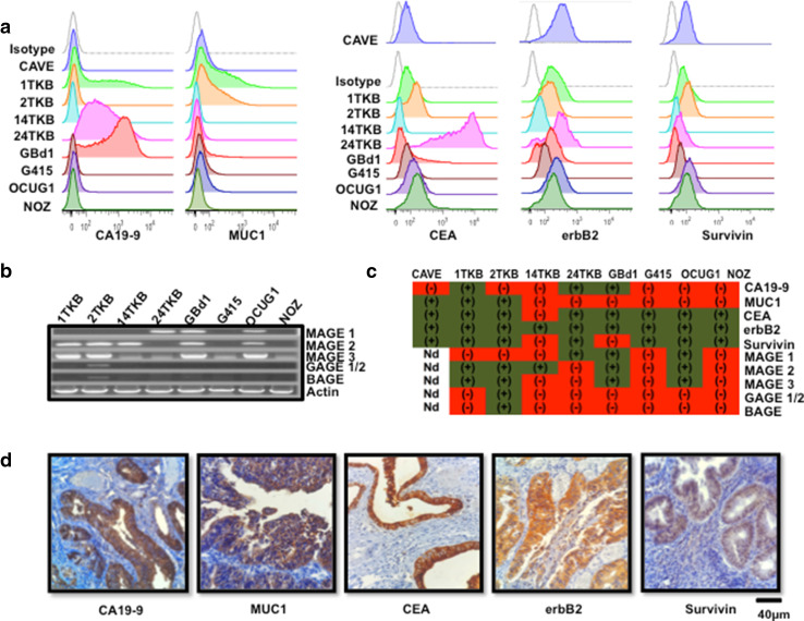
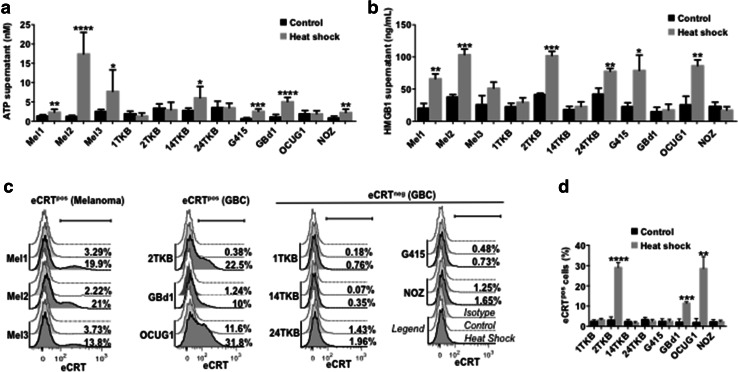
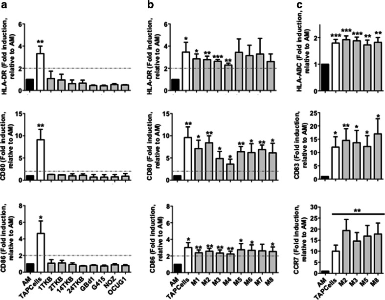
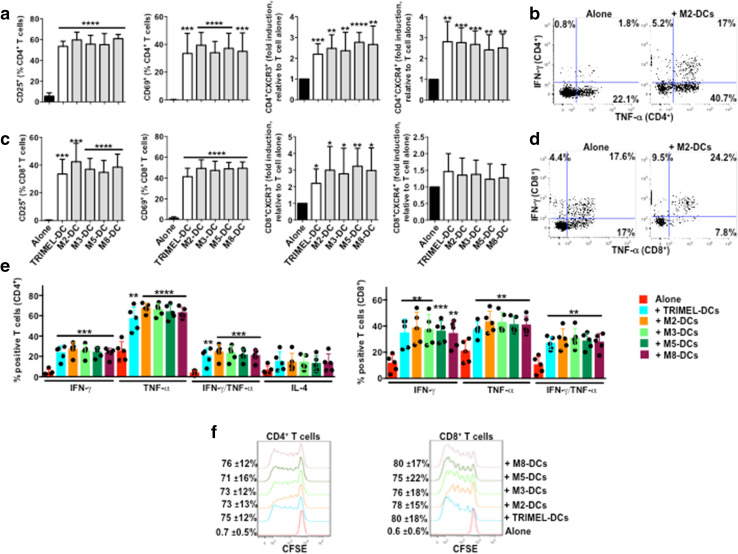
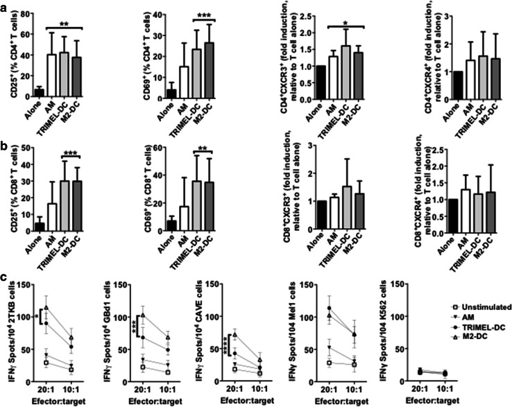
Immunotherapy based on checkpoint blockers has proven survival benefits in patients with melanoma and other malignancies. Nevertheless, a significant proportion of treated patients remains refractory, suggesting that in combination with active immunizations, such as cancer vaccines, they could be helpful to improve response rates. During the last decade, we have used dendritic cell (DC) based vaccines where DCs loaded with an allogeneic heat-conditioned melanoma cell lysate were tested in a series of clinical trials. In these studies, 60% of stage IV melanoma DC-treated patients showed immunological responses correlating with improved survival. Further studies showed that an essential part of the clinical efficacy was associated with the use of conditioned lysates. Gallbladder cancer (GBC) is a high-incidence malignancy in South America. Here, we evaluated the feasibility of producing effective DCs using heat-conditioned cell lysates derived from gallbladder cancer cell lines (GBCCL). By characterizing nine different GBCCLs and several fresh tumor tissues, we found that they expressed some tumor-associated antigens such as CEA, MUC-1, CA19-9, Erb2, Survivin, and several carcinoembryonic antigens. Moreover, heat-shock treatment of GBCCLs induced calreticulin translocation and release of HMGB1 and ATP, both known to act as danger signals. Monocytes stimulated with combinations of conditioned lysates exhibited a potent increase of DC-maturation markers. Furthermore, conditioned lysate-matured DCs were capable of strongly inducing CD4+ and CD8+ T cell activation, in both allogeneic and autologous cell co-cultures. Finally, in vitro stimulated CD8+ T cells recognize HLA-matched GBCCLs. In summary, GBC cell lysate-loaded DCs may be considered for future immunotherapy approaches.

Keywords: CITIM 2017; Dendritic cells; Gallbladder cancer; Immunotherapy; Melanoma; Tumor lysates.


Conflict of interest statement

The authors declare that they have no conflict of interest.

Figures

Fig. 1
Fig. 1
Tumor associated antigen expression in GBCCL and GBC fresh tumor samples. a Representative histograms for CA19-9, MUC-1, CEA, erbB2 and survivin expression in GBCCL evaluated by flow cytometry. Grey histograms indicate isotype control staining. b mRNA expression profiles for MAGE 1, 2, 3, GAGE 1/2 and BAGE in the GBCCL analyzed by RT-PCR. Actin was used as a housekeeping gene control. c Summary of tumor associated antigen expression in GBCCL. Green and red refers to positive or negative expression, respectively. ND not determined. d Representative photomicrographs of immunohistochemical staining for CA19-9, MUC-1, CEA, erbB2 and survivin in paraffin-embedded tumor biopsies obtained from Chilean GBC patients (scale bar, 40 µm)
Fig. 2
Fig. 2
Heat shock conditioning induces DAMP production in GBCCL. The levels of ATP (a) or HMGB1 (b) were evaluated in the supernatants from heat shock-treated or control cells. c Representative histograms showing the extracellular expression levels of translocated calreticulin (eCRT) in heat shock-treated (dark grey) or control (light grey) melanoma and GBC cells. White histograms indicate isotype control staining. The percentage of eCRT positive (eCRTpos) for each condition is shown. d Statistical analysis of eCRT translocation induced by heat shock in GBCCL. Bars represent averages and standard deviations of three (bd) or five-seven (a) measurements of three independent experiments. *p < 0.05; **p < 0.01; ***p < 0.001; ****p < 0.0001
Fig. 3
Fig. 3
Heat shock-conditioned GBCCL lysate mixtures, but not lysates from individual cell lines, induce differentiation of activated monocytes into mature DCs. Surface expression of HLA-DR, CD80, CD86 (a, b), and HLA-ABC, CD83, and CCR7 (c) were evaluated by flow cytometry on activated monocytes (AM) incubated or not for 24 h with 100 µg/mL of heat shock-conditioned tumor lysates generated from individual GBCCL (a) or mixtures (M1-M8) of three different GBCCL (b, c). Bars represent the average and SD of the fold induction of the integrated MFI (iMFI: % positive cells × geoMFI of positive cells) for each marker relative to AM from at least three independent experiments. Evaluated cell lysates mix were made as follows: M1 (24TKB + GBd1 + G415); M2 (2TKB + 24TKB + GBd1); M3 (1TKB + 2TKB + 24TKB); M4 (OCUG1 + GBd1 + G415); M5 (2TKB + G415 + OCUG1); M6 (NOZ + OCUG 1 + G415); M7 (1TKB + 14TKB + 24TKB); and M8 (24TKB + OCUG1 + G415). *p < 0.05; **p < 0.01; ***p < 0.001
Fig. 4
Fig. 4
Activation of allogeneic T cells by monocyte-derived DCs matured with different heat shock-conditioned GBC lysates. Purified CD3+ T cells were co-cultured for 5 days with allogeneic TRIMEL-, M2-, M3-, M5-, M8-DCs or without DCs. The surface expression of CD25, CD69, CXCR3 and CXCR4 (a), the intracellular levels of IFN-γ, TNF-α and IL-4 (bd), and proliferation (e) were evaluated in the CD4+ and CD8+ T cells populations by flow cytometry. a, d Bars represent the average and SD from five independent experiments of the % of T cells positive for each marker, with the exception of CXCR3 and CXCR4 data that are shown as fold induction of the MFI relative to unstimulated T cells. Representative dot plots of IFN-γ and TNF-α production in allogeneic CD4+ (b) and CD8+ (c) T cells co-cultured with M2-DCs. e The percentage and SD of proliferating T cells are showed on the left of each histograms. Evaluated cell lysates mix were made as follows: M2 (2TKB + 24TKB + GBd1); M3 (1TKB + 2TKB + 24TKB); M5 (2TKB + G415 + OCUG1); and M8 (24TKB + OCUG1 + G415). *p < 0.05; **p < 0.01; ***p < 0.001; ****p < 0.0001 (comparison versus unstimulated T cells)
Fig. 5
Fig. 5
T cells activated by autologous monocyte-derived DCs loaded with a heat shock conditioned GBC lysate recognize HLA-A2-matched GBCCL. ac Purified CD3+ T cells were co-cultured for 14 days with autologous HLA-A2+ AM, TRIMEL-DCs, M2-DCs or cultured alone. The surface expression of CD25, CD69, CXCR3 and CXCR4 (a, b) were evaluated in the CD4+ (a) and CD8+ (b) T cells populations by flow cytometry. Bars represent the average and SD from at least three independent experiments of the % of T cells positive for each marker, with the exception of CXCR3 and CXCR4 data that are shown as fold induction of the MFI relative to unstimulated T cells. *p < 0.05; **p < 0.01; ***p < 0.001 (comparison versus unstimulated T cells). c Sorted CD8+ T cells were challenged for 16 h with the HLA-A2+ GBCCL 2TKB, GBd1, CAVE, the melanoma cell line Mel1 or K562 cells. IFN-γ release was measured by ELISPOT at different effector:target ratios as indicated. Data represent the average and SD of at least three independent experiments. *p < 0.05; ***p < 0.001; ****p < 0.0001 (comparison M2-DC versus TRIMEL-DCs stimulated T cells). M2 refer to the mixture made from three different GBCCL

References

    1. Rojas-Sepúlveda D, Gleisner MA, Pereda C, López MN, Salazar-Onfray F. Tumor cell lysates as maturation stimulus and antigen source for therapeutic dendritic cells against gallbladder cancer. Eur J Immunol. 2016;46(S1):1221–1222.
    1. Rojas-Sepúlveda D, Gleisner MA, Pereda C, López MN, Salazar-Onfray F. Tumor lysate loaded dendritic cells induce a T cell specific antitumor response against gallbladder cancer. J Immunol. 2017;198(1 Supplement):79.18.
    1. Guida M, Pisconte S, Colucci G. Metastatic melanoma: the new era of targeted therapy. Expert Opin Ther Targets. 2012;16 Suppl 2:S61–S70. doi: 10.1517/14728222.2011.645807. - DOI - PubMed
    1. Larkin J, Chiarion-Sileni V, Gonzalez R, Grob JJ, Cowey CL, Lao CD, et al. Combined nivolumab and ipilimumab or monotherapy in untreated melanoma. N Engl J Med. 2015;373:23–34. doi: 10.1056/NEJMoa1504030. - DOI - PMC - PubMed
    1. Farolfi A, Ridolfi L, Guidoboni M, Nicoletti SV, Piciucchi S, Valmorri L, et al. Ipilimumab in advanced melanoma: reports of long-lasting responses. Melanoma Res. 2012;22:263–270. doi: 10.1097/CMR.0b013e328353e65c. - DOI - PubMed

MeSH terms