Immunolocalization of Surfactant Proteins SP-A, SP-B, SP-C, and SP-D in Infantile Labial Glands and Mucosa
- PMID: 29601229
- PMCID: PMC6055263
- DOI: 10.1369/0022155418766063
Immunolocalization of Surfactant Proteins SP-A, SP-B, SP-C, and SP-D in Infantile Labial Glands and Mucosa
Abstract
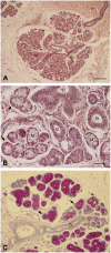
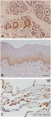
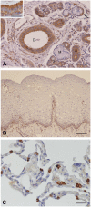
Surfactant proteins in different glandular structures of the oral cavity display antimicrobial activity for protection of invading microorganisms. Moreover, they are involved in lowering liquid tension in fluids and facilitate secretion flows. Numerous investigations for studying the occurrence of surfactant proteins in glandular tissues were performed using different methods. In the oral cavity, minor salivary glands secrete saliva continuously for the maintenance of a healthy oral environment. For the first time, we could show that infantile labial glands show expression of the surfactant proteins (SP) SP-A, SP-B, SP-C, and SP-D in acinar cells and the duct system in different intensities. The stratified squamous epithelium of the oral mucosa revealed positive staining for SPs in various cell layers.
Keywords: human labial gland; immunohistochemistry; minor salivary gland; surfactant proteins.
Conflict of interest statement
Figures


References
-
- Liley HG, Ertsey R, Gonzales LW, Odom MW, Hawgood S, Dobbs LG, Ballard PL. Synthesis of surfactant components by cultured type II cells from human lung. Biochim Biophys Acta. 1988;961(1):86–95. - PubMed
-
- Goerke J. Pulmonary surfactant: functions and molecular composition. Biochim Biophys Acta. 1998;1408(2–3):79–89. - PubMed
-
- McCormack FX. Structure, processing and properties of surfactant protein A. Biochim Biophys Acta. 1998;1408(2–3):109–31. - PubMed
-
- Pikaar JC, Voorhout WF, van Golde LM, Verhoef J, van Strijp JA, van Iwaarden JF. Opsonic activities of surfactant proteins A and D in phagocytosis of gram-negative bacteria by alveolar macrophages. J Infect Dis. 1995;172(2):481–9. - PubMed
-
- Mariencheck WI, Savov J, Dong Q, Tino MJ, Wright JR. Surfactant protein A enhances alveolar macrophage phagocytosis of a live, mucoid strain of P. aeruginosa. Am J Physiol. 1999;277(4, Pt 1):L777–86. - PubMed
MeSH terms
Substances
LinkOut - more resources
Full Text Sources
Other Literature Sources

