Review
. 2018 Mar 30;10(4):161.
doi: 10.3390/v10040161.

Invertebrate Iridoviruses: A Glance over the Last Decade

Affiliations
Review

Invertebrate Iridoviruses: A Glance over the Last Decade

İkbal Agah İnce et al. Viruses. .

Abstract

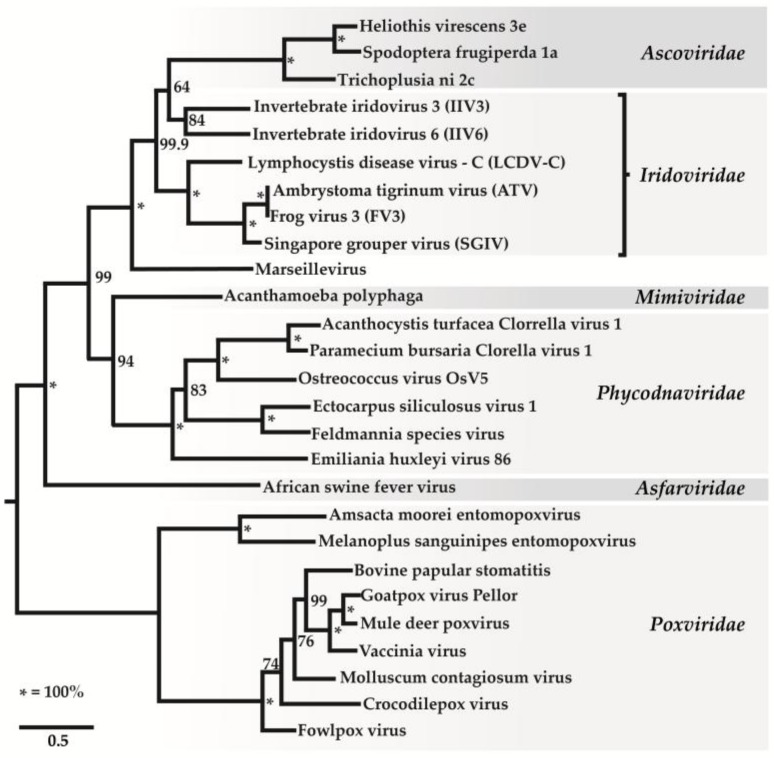
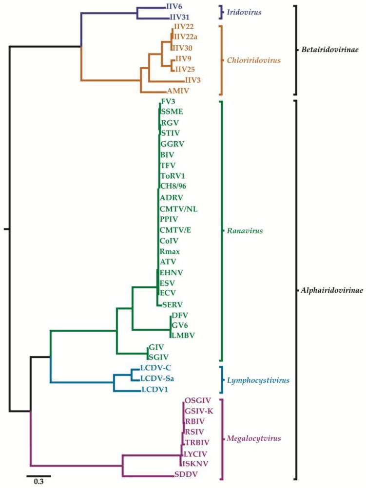
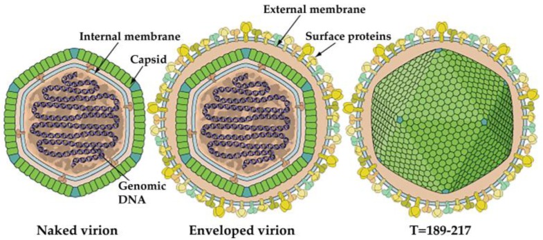
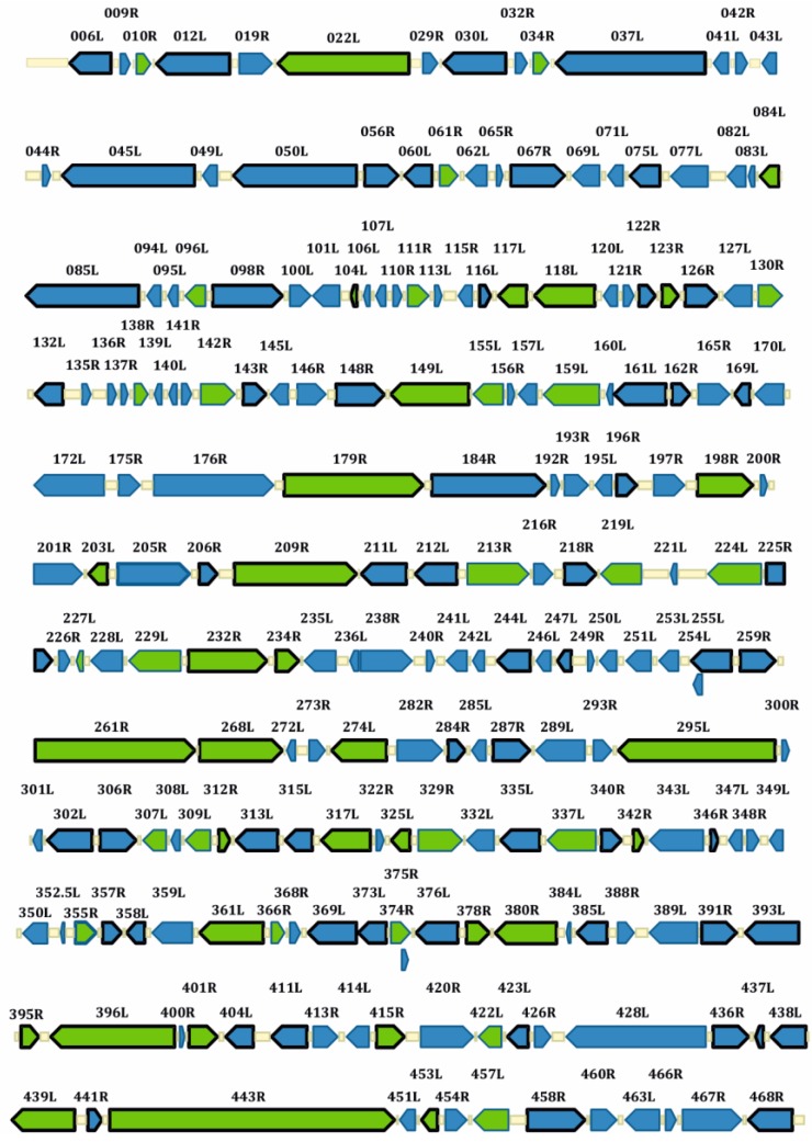
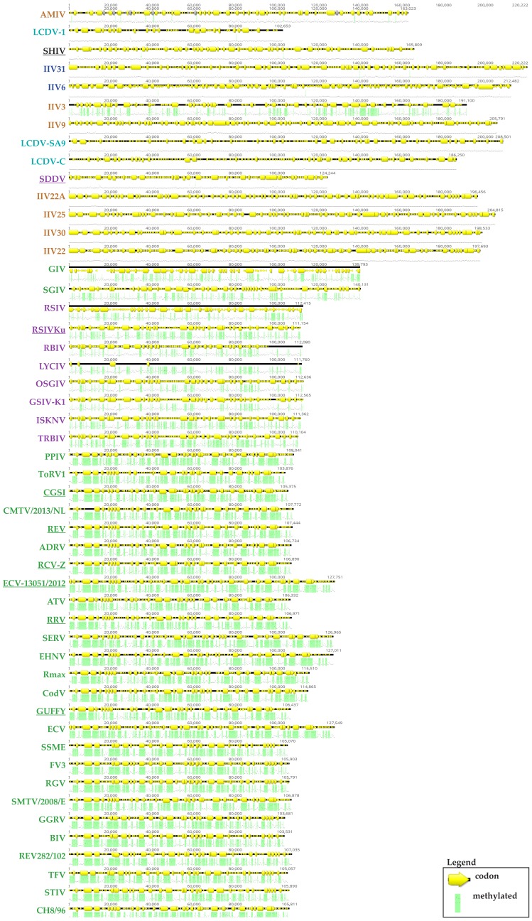
Members of the family Iridoviridae (iridovirids) are large dsDNA viruses that infect both invertebrate and vertebrate ectotherms and whose symptoms range in severity from minor reductions in host fitness to systemic disease and large-scale mortality. Several characteristics have been useful for classifying iridoviruses; however, novel strains are continuously being discovered and, in many cases, reliable classification has been challenging. Further impeding classification, invertebrate iridoviruses (IIVs) can occasionally infect vertebrates; thus, host range is often not a useful criterion for classification. In this review, we discuss the current classification of iridovirids, focusing on genomic and structural features that distinguish vertebrate and invertebrate iridovirids and viral factors linked to host interactions in IIV6 (Invertebrate iridescent virus 6). In addition, we show for the first time how complete genome sequences of viral isolates can be leveraged to improve classification of new iridovirid isolates and resolve ambiguous relations. Improved classification of the iridoviruses may facilitate the identification of genus-specific virulence factors linked with diverse host phenotypes and host interactions.

Keywords: classification; genomics; invertebrate iridoviruses; proteomics.


Conflict of interest statement

The authors declare no conflict of interest.

Figures

Figure 1
Figure 1
Phylogenetic relationships of iridovirids with other nucleocytoplasmic large DNA viruses A maximum-likelihood tree constructed based on concatenated alignments of 1849 positions of five NCLDV core proteins: A1L/VLTF2 transcription factor, A32 ATPase, D5 type ATPase, DNA polymerase B, and major capsid protein using the TreeFinder program with the estimated site rates heterogeneity and with the WAG (Whelan and Goldman) substitution model. The expected-likelihood weights of 1000 local rearrangements were used as confidence values of TreeFinder tree branches. Scale bar represents amino acid substitutions per site (This figure was used with permission from PNAS [8] and ICTV [4] where it was presented earlier and slightly modified its presentation).
Figure 2
Figure 2
ML analysis based on 26 core genes conserved among all members of the family Iridoviridae (iridovirids). The phylogram shows a clear division of genera within the Alphairidovirinae (Ranavirus, green; Lymphocystivirus, cyan; Megalocytivirus, purple) and Betairidovirinae (Iridovirus, blue; Chloriridovirus, brown) subfamilies. Virus species and isolates are indicated by abbreviations, listed in Table 2. Members of accepted species are shown in bold typeface. The tree was constructed using maximum likelihood analysis using IQ-TREE open source software [9] under optimum substitution model LG+I+G4 according to Bayesian Information Criterion (BIC) [10].and the concatenated amino acid (aa) sequences of 26 core genes (19,773 aa characters including gaps) from 45 completely sequenced iridovirus genomes. The tree was midpoint rooted and branch lengths are based on the number of inferred substitutions, as indicated by the scale bar. The branch points that separate the iridovirid genera are supported by bootstrap values greater than 99%. For other branch points all bootstrap values are >70% except for those displaying high levels of amino acid similarity, e.g., TFV vs. BIV/GGRV, 66%; PPIV vs. CMTV/2013/NL et al., 49%; TRBIV vs. RSIV et al., 57%; and LYCIV vs. TRBIV et al., 56%. Scale bar represents amino acid substitutions per site. This figure was published as an ICTV report [1,2].
Figure 3
Figure 3
Graphical representation of the iridovirid structure [73]. Source: ViralZone: www.expasy.org/viralzone, SIB Swiss Institute of Bioinformatics.
Figure 4
Figure 4
IIV-6 linear genomic map presenting virion and viral proteins identified in infected cells using mass spectrometry (genome size 212,482 bp). Arrows represent the transcriptional direction of the ORFs. All arrows with a black frame represent ORFs for which the corresponding protein was detected in the infected-cell proteomic analysis [127]. Green arrows represent known virion proteins [128]. This figure was published previously [127].
Figure 5
Figure 5
In silico prediction of methylation sites of 50 iridovirid genomes. The Geneious CpG Plugin (Biomatters Ltd., Auckland, New Zealand) predicts likely methylation sites according to the Hidden Markov models described by Richard Durbin. Green indicates potential methylation sites and yellow arrows indicates viral coding region of a genes. Methylation patterns IIVs versus VIVs could be distinguished, with the exceptions of IIV3 from IIVs and SDDV from SDDV as well as LCDV-1.
Figure 6
Figure 6
Schematic representation of iridoviral replication. This figure was obtained from the 10th report of ICTV with permission [1,2].

References

    1. Chinchar V.G., Hick P., Ince I.A., Jancovich J.K., Marschang R., Qin Q., Subramaniam K., Waltzek T.B., Whittington R., Williams T., et al. ICTV Virus Taxonomy Profile: Iridoviridae. J. Gen. Virol. 2017;98:890–891. doi: 10.1099/jgv.0.000818. - DOI - PMC - PubMed
    1. Chinchar V.G., Waltzek T.B., Subramaniam K. Ranaviruses and other members of the family Iridoviridae: Their place in the virosphere. Virology. 2017;511:259–271. doi: 10.1016/j.virol.2017.06.007. - DOI - PubMed
    1. Chinchar V., Essbauer S., He J., Hyatt A., Miyazaki T., Seligy V., Williams T. Virus Taxonomy Eighth Report of the International Committee on Taxonomy of Viruses. Elsevier Academic Press; San Diego, CA, USA: 2005. Family Iridoviridae; pp. 145–162.
    1. King A.M., Lefkowitz E., Adams M.J., Carstens E.B. Virus Taxonomy: Ninth Report of the International Committee on Taxonomy of Viruses. Elsevier; Amsterdam, The Netherlands: 2011.
    1. Xeros N. A second virus disease of the leatherjacket, Tipula paludosa. Nature. 1954;174:562–563. doi: 10.1038/174562a0. - DOI

LinkOut - more resources