. 2018 Mar 30;13(3):e0195335.
doi: 10.1371/journal.pone.0195335. eCollection 2018.

HMGB1/IL-1β complexes in plasma microvesicles modulate immune responses to burn injury

Affiliations

HMGB1/IL-1β complexes in plasma microvesicles modulate immune responses to burn injury

Leon G Coleman Jr et al. PLoS One. .

Abstract

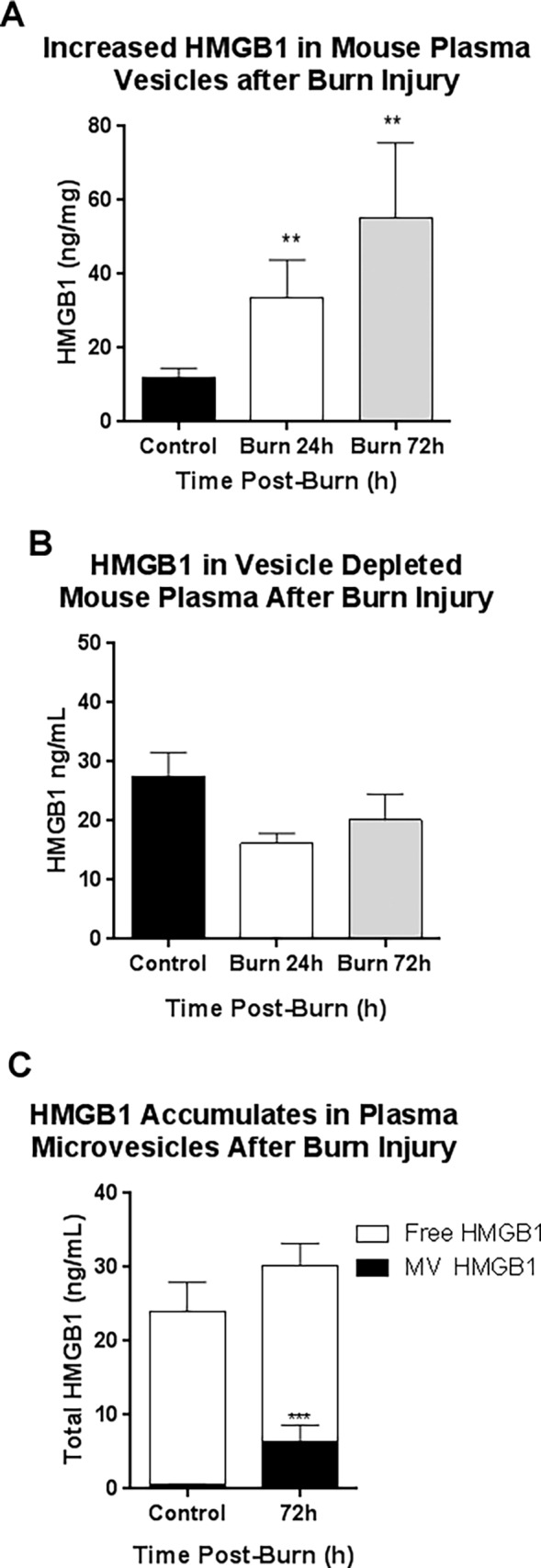
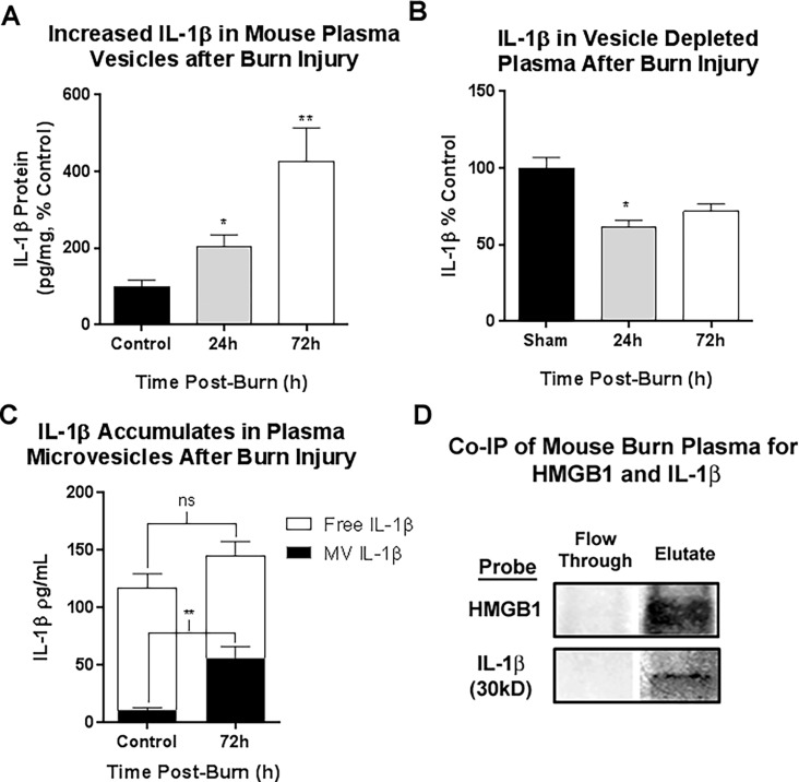
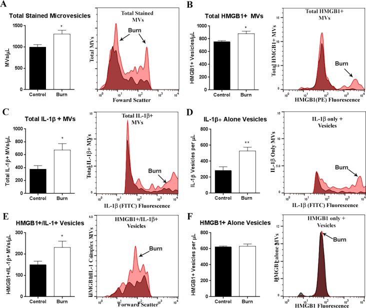
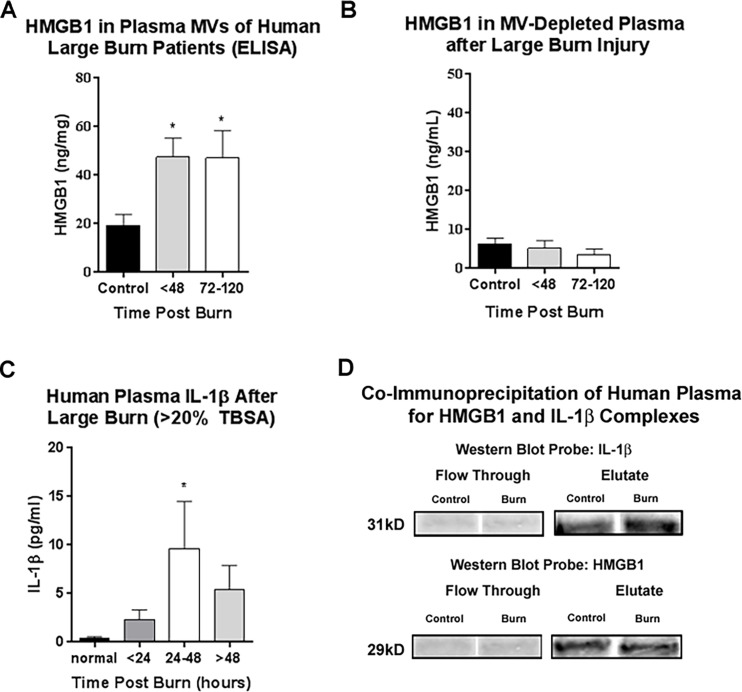
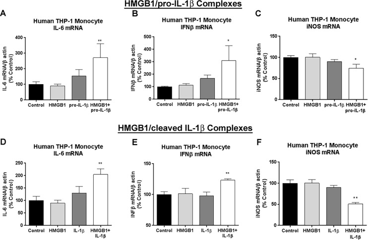
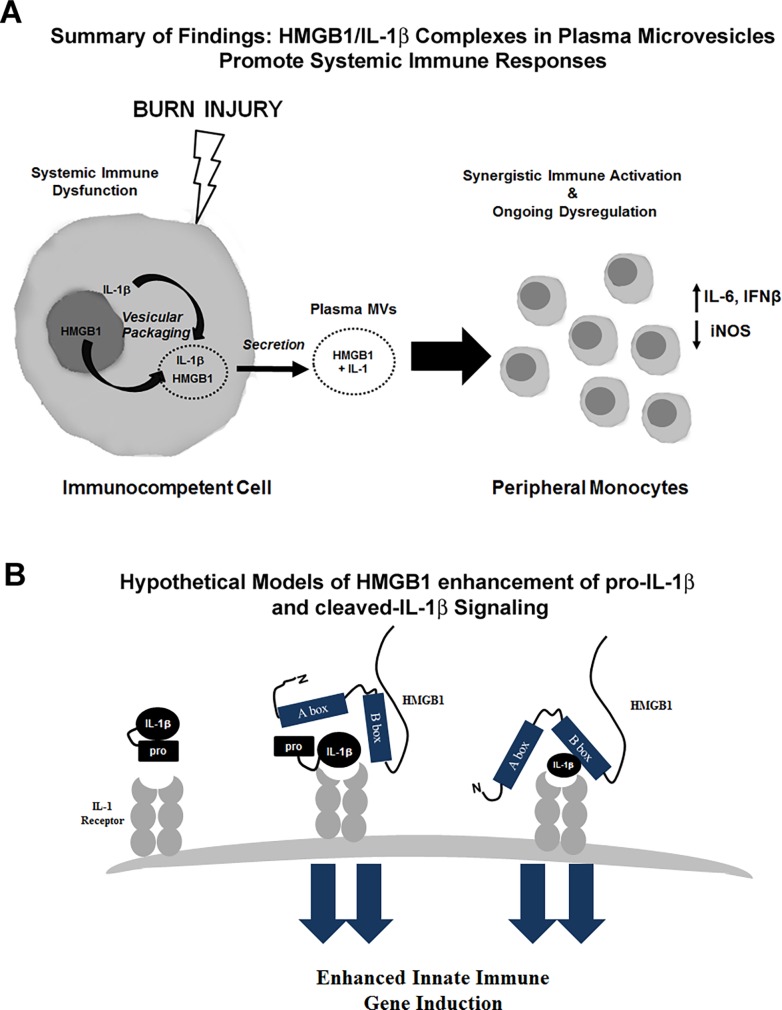
Modulating immune responses to sepsis and trauma remain one of the most difficult challenges in modern medicine. Large burn injuries (LBI) are a severe form of trauma associated with sepsis, immune impairment, and mortality. Immune dysfunction after LBI is complex, involving both enhanced and impaired immune activation. The release of Damage-Associated Molecular Patterns (DAMPs), such as HMGB1, and cytokines (e.g. IL-1β) creates an environment of immune dysfunction often leading to end organ failure and death. Both HMGB1 and IL-1β have been found to play critical roles in sepsis and post-burn immune dysfunction. HMGB1 and IL-1β have been shown previously to form potent complexes in vitro. We recently identified the presence of HMGB1/IL-1β heterocomplexes in human tissue. We now find HMGB1/IL-1β complexes in human and mouse plasma, and identify a synergistic role of HMGB1/IL-1β complexes in post-burn immune dysfunction. In both humans and mice, we found that HMGB1 was enriched in plasma microvesicles (MVs) after LBI. HMGB1 was found form complexes with IL-1β. Using flow cytometry of mouse plasma MVs, we identified an increase in an HMGB1+/IL-1β+ MVs. Using co-IP, HMGB1 was found to bind the pro-form of IL-1β in mouse and human plasma. Pro-IL-1β, which is traditionally considered inactive, became active when complexed with HMGB1. Human THP-1 monocytes treated with HMGB1-pro-IL-1β complexes showed increased transcription of LBI associated cytokines IL-6 and IFNβ along with suppression of iNOS, mimicking findings associated with LBI. These findings identify that HMGB1/IL-1β complexes released after burn injuries can modulate immune responses, and microvesicles are identified as a novel reservoir for these immune mediators. These complexes might serve as novel immune targets for the treatment of systemic immune responses due to LBI or other causes of sepsis.


Conflict of interest statement

Competing Interests: The authors have declared that no competing interests exist.

Figures

Fig 1
Fig 1. HMGB1 is concentrated in mouse plasma microvesicles (MVs), but not vesicle depleted plasma up to 72 hours after burn injury.
Mice underwent a 20% TBSA thermal injury and were sacrificed at either 24 or 72 hours post-burn. Plasma microvesicles (MVs) were isolated from by sequential centrifugation. HMGB1 levels were assessed by ELISA in the MVs and MV-depleted plasma. (A) HMGB1 was increased in plasma MVs up to 4.6-fold after burn injury. This was observed at 24 hours post-burn (33.6±10.2 vs. 12.06±2.43 ng/mg total protein, mean±SEM, Burn vs Control, **p<0.01,) and at 72 hours post-burn (55.35±20.23 vs. 12.06±2.43 ng/mg total protein, Burn vs Control, mean±SEM, **p<0.01) N = 13 control and 5–6 burn mice per group. (B) HMGB1 levels were not increased in the MV-depleted plasma (C) Analysis of plasma fraction, MV versus MV free plasma, showed that plasma HMGB1 increases at 72 hours were due to increases in MV fraction.
Fig 2
Fig 2. IL-1β is concentrated in mouse plasma microvesicles (MVs), but not vesicle depleted plasma up to 72 hours after burn injury.
Mice underwent a 20% TBSA thermal injury and were sacrificed at either 24 or 72 hours post-burn. Plasma microvesicles (MVs) were isolated from by sequential centrifugation. IL-1β levels were assessed by ELISA in the MVs and MV-depleted plasma. (A) IL-1β was increased in plasma MVs up to 426% of control levels. At 24 hours post-burn IL-1β was 2-fold control levels; 203.1±32.1 vs. 100±10.7% control, mean±SEM, Burn vs Control, group *p<0.05, N = 3 control, 4 burn. At 72 hours post-burn IL-1β levels in MVs were 400% control values; 426.1±88.09 vs. 100 ± 10.7% control, Burn vs Control, mean±SEM, **p<0.01, N = 5 per group. Data from two separate experiments were combined and presented as percent control. (B) In MV-depleted plasma, IL-1β levels were not increased. At 24 hours, MV-free IL-1β was significantly reduced by 21%; 100±8.43% vs 79.12±4.02%, mean±SEM, Control vs Burn, p<0.05, N = 4–5. At 72 hours post burn a nonsignificant reduction in IL-1β was observed; 100±10.10% vs 80.43±12.36%, mean±SEM, Control vs Burn, p = 0.24, N = 5–7. (C) Analysis of plasma fraction, MV versus MV free plasma, showed that plasma IL-1β increases at 72 hours were due to increases in MV fraction. (D) Co-immunoprecipitation (Co-IP) of mouse plasma for HMGB1 was performed. Column flow through and eluate were assessed by western blot for HMGB1 and IL-1β. Both HMGB1 and IL-1β were found in the eluate indicating HMGB1 and IL-1β complex formation in mouse plasma. Both the 31kD pro-form of IL-1β and HMGB1 were detected in the eluate, but not the flow through demonstrating the presence of HMGB1/pro-IL-1β complexes in human plasma.
Fig 3
Fig 3. HMGB1 and IL-1β Co-localize in plasma microvesicles (MVs) following burn injury.
Mice underwent a 20% TBSA thermal injury and were sacrificed at 72 hours post-burn. Plasma microvesicles (MVs) were isolated from by sequential centrifugation. Vesicles were permeabilized using Fix/Perm buffer and labeled with anti-HMGB1 and anti-IL-1β antibodies. MVs between 0.1–1.0μm were identified using MegaMix size gating beads. Positive staining was differentiated from background by comparing to single color controls for each antibody. (A) The total number of stained MVs was increased by 34% at 72 hours after burn injury; **p<0.01, N = 8 control, 6 burn. (B) The total number of HMGB1+ MVs was increased by 17%; 753.5 ± 19.6 vs 879.1 ± 41.7; Control vs Burn N = 7 Control, 6 Burn (C) The total number of IL-1+MVs was increased by 78%; 377.7 ± 54.9 vs 672.5 ± 99.1 Control vs Burn N = 8 Control, 6 Burn. (D) MVs positive for IL-1β alone were increased after burn injury by 61%; 281.9 ± 51.22 vs 536.6 ± 45.76, Control vs Burn, *p<0.05, N = 7 control, 5 burn. (E) MVs positive for both HMGB1 and IL-1β were increased by 64%; 145.4 ± 15.30 vs 237.8 ± 32.74, Control vs Burn, *p<0.05, N = 7 control, 6 burn. (F) MVs positive for HMGB1 alone were not changed after burn injury, 612.7 ± 13.27 vs 641.3 ± 22.73, Control vs Burn, p = 0.27, N = 8 control, N = 6 burn. Representative relative frequency histograms depicting different MV populations for each panel are included: red-burn, white-control. **p<0.01, p<0.05, t-test vs control.
Fig 4
Fig 4. Human large burn injuries cause increased HMGB1 release in microvesicles (MVs), IL-β, and HMGB1/IL-1 complexes in plasma.
In two different patient groups of adult patients admitted to the North Carolina Jaycee Burn Center at the University of North Carolina at Chapel Hill hospital were enrolled into the study and serial blood draws were taken. IL-1β and HMGB1 were measured in plasma and plasma MVs respectively. (A-B) Plasma was collected from 15 human burn patients with a mean age of 36.5 (range 12–77); 80% male, mean %TBSA 14.5% (range 1.5–42%). Plasma collections were obtained during the first 48 hours of admission and between 72–120 hours after admission. See Table 2 for details. (A) Microvesicles (MVs) were isolated by centrifugation. HMGB1 was measured in MVs by ELISA. HMGB1 was increased in plasma MVs 2.5-fold after burn injury. This was observed both within 48 hours (47.6±7.73 vs. 19.32±4.5 ng/mg total protein, Burn vs Control) and at 72–120 hours after admission (47.09±19.5 vs. 19.32±4.5 ng/mg total protein, Burn vs Control, mean±SEM, *p<0.05) N = 3–6 per group. (B) HMGB1 was measured in MV-depleted plasma by ELISA. HMGB1 in MV-depleted plasma was unchanged after burn injury. (C-D) Plasma was collected from 22 patients, mean age 42 (range 18–78); 82% male, mean %TBSA 26% (range 5–45%), see Table 3. The first blood sample was obtained at 0–24 hours following initial burn injury, the second was obtained at 24–48 hours following injury, and the third was obtained at 48–72 hours following injury. (C) Large burn (>20% TBSA, n = 8) was associated with a significant (*p < 0.05) increase in IL-1β plasma concentration at 24–48 hours compared to control, normal individuals. (D) Co-immunoprecipitation was performed in human plasma to confirm the presence of HMGB1/IL-1β complexes. Western blot was performed and the flow through and eluate probed for IL-1β and HMGB1. Both the 31kD pro-form of IL-1β and HMGB1 were detected in the eluate, but not the flow through demonstrating the presence of HMGB1/pro-IL-1β complexes in human plasma.
Fig 5
Fig 5. Enhanced induction of proinflammatory gene expression by HMGB1/IL-1β complex.
Human THP-1 monocytes were incubated with either recombinant (r) human HMGB1 (4nM, 100ng/mL), recombinant pro-IL-1β (rpro-IL-1β, 4nM) or rHMGB1/r pro-IL-1β complexes. Cell lysates were harvested 24 hours later and analyzed by RT-PCR for expression of immune gene of interest. (A) Neither HMGB1 nor pro-IL-1β alone significantly altered IL-6 mRNA. However, rHMGB1/r pro-IL-1β complexes caused a 2.7-fold increase in IL-6 gene expression (B) Neither HMGB1 nor pro-IL-1β alone significantly altered IFNβ mRNA. rHMGB1/r pro-IL-1β complexes caused a 3.1-fold increase in IFNβ gene expression (C) rHMGB1/rpro-IL-1β caused a 36% reduction in iNOS gene expression, while neither HMGB1 nor pro-IL-1β alone caused changes (D) r-pro-IL-1β caused a 2.5-fold increase in NOX-2 alone. rHMGB1/rIL-1β complexes showed no further increase in NOX-2 gene expression. ***p<0.001, **p<0.01, *p<0.05 vs control. N = 2–6 culture wells per group.
Fig 6
Fig 6. Proposed model of enhanced activation by HMGB1/IL-1β complexes.
(A) Schematic illustrating secretion of HMGB1 and IL-1 after burn injury. HMGB1 translocates from the nucleus to the cytoplasm where it is packaged in vesicles. HMGB1 and IL-1β were increased in plasma MVs after burn injury. HMGB1/IL-1β complexes caused enhanced increases in IL-6 and IFNβ gene induction in human THP-1 monocytes. (B) Hypothetical models of enhanced immune activation by HMGB1/pro-IL-1β and HMGB1/cleaved-IL-1β complexes. Pro-IL-1β is considered inactive at the IL-1 receptor. When coupled with HMGB1, pro-IL-1β caused enhanced immune activation in a manner similar to HMGB1/cleaved IL-1β complexes.

References

    1. Yang H, Ochani M, Li J, Qiang X, Tanovic M, Harris HE, et al. Reversing established sepsis with antagonists of endogenous high-mobility group box 1. Proceedings of the National Academy of Sciences of the United States of America. 2004;101(1):296–301. doi: 10.1073/pnas.2434651100 ; PubMed Central PMCID: PMC314179. - DOI - PMC - PubMed
    1. Lamkanfi M, Sarkar A, Vande Walle L, Vitari AC, Amer AO, Wewers MD, et al. Inflammasome-dependent release of the alarmin HMGB1 in endotoxemia. Journal of immunology. 2010;185(7):4385–92. doi: 10.4049/jimmunol.1000803 ; PubMed Central PMCID: PMCPMC3428148. - DOI - PMC - PubMed
    1. Wang H, Bloom O, Zhang M, Vishnubhakat JM, Ombrellino M, Che J, et al. HMG-1 as a late mediator of endotoxin lethality in mice. Science. 1999;285(5425):248–51. . - PubMed
    1. Opal SM, Fisher CJ Jr., Dhainaut JF, Vincent JL, Brase R, Lowry SF, et al. Confirmatory interleukin-1 receptor antagonist trial in severe sepsis: a phase III, randomized, double-blind, placebo-controlled, multicenter trial. The Interleukin-1 Receptor Antagonist Sepsis Investigator Group. Critical care medicine. 1997;25(7):1115–24. . - PubMed
    1. Fisher CJ Jr., Dhainaut JF, Opal SM, Pribble JP, Balk RA, Slotman GJ, et al. Recombinant human interleukin 1 receptor antagonist in the treatment of patients with sepsis syndrome. Results from a randomized, double-blind, placebo-controlled trial. Phase III rhIL-1ra Sepsis Syndrome Study Group. JAMA. 1994;271(23):1836–43. . - PubMed

Publication types