. 2018 Sep;32(9):4941-4954.
doi: 10.1096/fj.201701455RR. Epub 2018 Mar 30.

Cardiac fibroblast-specific p38α MAP kinase promotes cardiac hypertrophy via a putative paracrine interleukin-6 signaling mechanism

Affiliations

Cardiac fibroblast-specific p38α MAP kinase promotes cardiac hypertrophy via a putative paracrine interleukin-6 signaling mechanism

Sumia A Bageghni et al. FASEB J. 2018 Sep.

Abstract

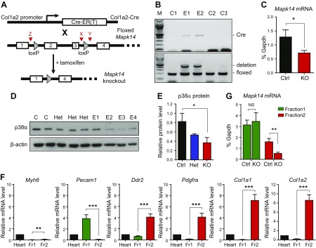
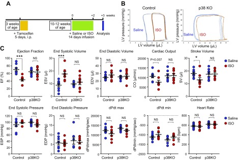
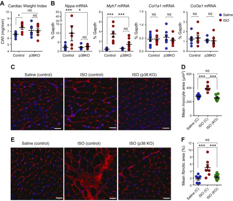
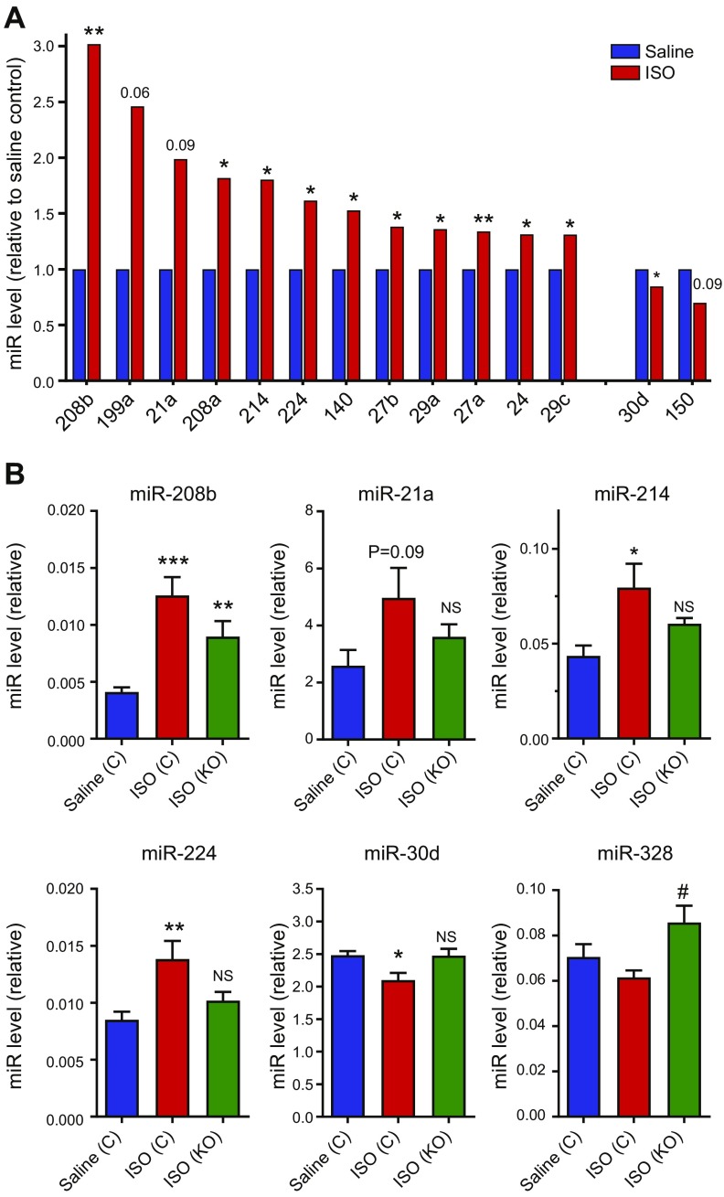
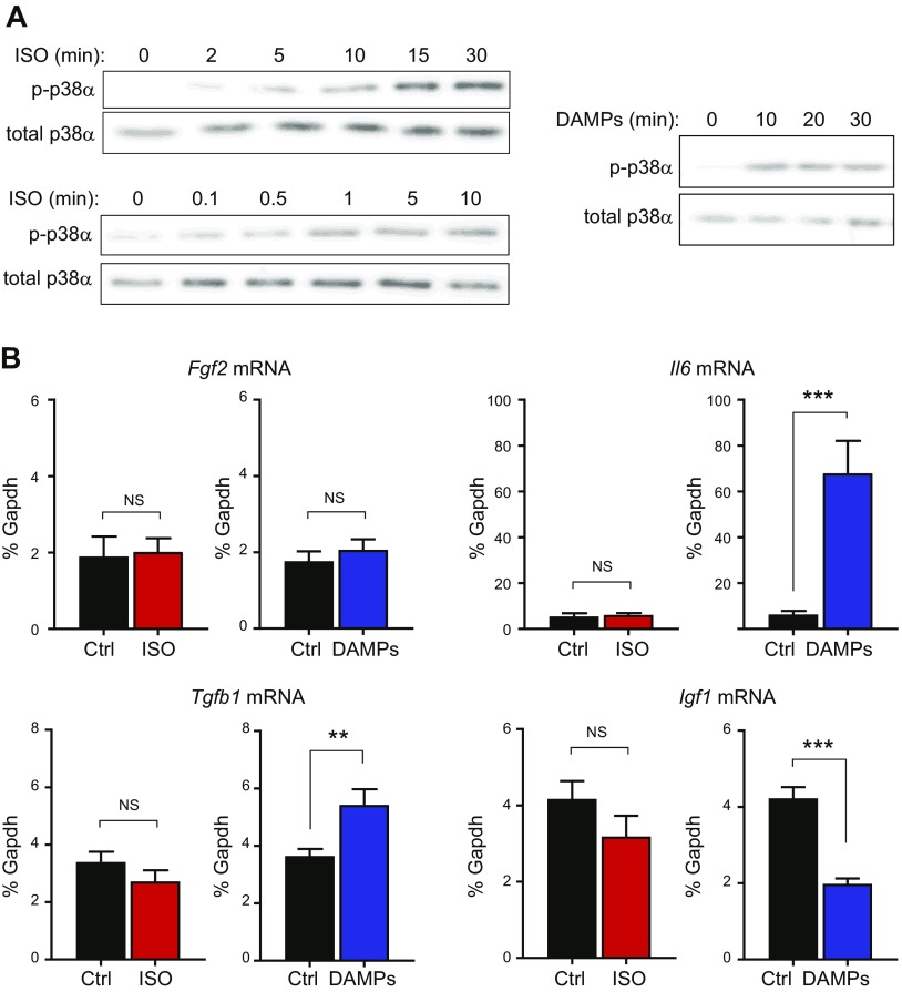
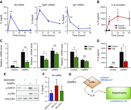
Recent studies suggest that cardiac fibroblast-specific p38α MAPK contributes to the development of cardiac hypertrophy, but the underlying mechanism is unknown. Our study used a novel fibroblast-specific, tamoxifen-inducible p38α knockout (KO) mouse line to characterize the role of fibroblast p38α in modulating cardiac hypertrophy, and we elucidated the mechanism. Myocardial injury was induced in tamoxifen-treated Cre-positive p38α KO mice or control littermates via chronic infusion of the β-adrenergic receptor agonist isoproterenol. Cardiac function was assessed by pressure-volume conductance catheter analysis and was evaluated for cardiac hypertrophy at tissue, cellular, and molecular levels. Isoproterenol infusion in control mice promoted overt cardiac hypertrophy and dysfunction (reduced ejection fraction, increased end systolic volume, increased cardiac weight index, increased cardiomyocyte area, increased fibrosis, and up-regulation of myocyte fetal genes and hypertrophy-associated microRNAs). Fibroblast-specific p38α KO mice exhibited marked protection against myocardial injury, with isoproterenol-induced alterations in cardiac function, histology, and molecular markers all being attenuated. In vitro mechanistic studies determined that cardiac fibroblasts responded to damaged myocardium by secreting several paracrine factors known to induce cardiomyocyte hypertrophy, including IL-6, whose secretion was dependent upon p38α activity. In conclusion, cardiac fibroblast p38α contributes to cardiomyocyte hypertrophy and cardiac dysfunction, potentially via a mechanism involving paracrine fibroblast-to-myocyte IL-6 signaling.-Bageghni, S. A., Hemmings, K. E., Zava, N., Denton, C. P., Porter, K. E., Ainscough, J. F. X., Drinkhill, M. J., Turner, N. A. Cardiac fibroblast-specific p38α MAP kinase promotes cardiac hypertrophy via a putative paracrine interleukin-6 signaling mechanism.

Keywords: cardiomyocyte; heart; isoproterenol; microRNA; mouse.


Conflict of interest statement

The authors thank Drs. Juan-Jose Ventura [Katholieke Universiteit (KU) Leuven, Leuven, Belgium] and Manolis Pasparakis (University of Cologne, Cologne, Germany), for the provision of floxed Mapk14 mice, and Emmanuel Pinteaux (University of Manchester, Manchester, United Kingdom) for the provision of floxed Il1r1 mice. This work was supported by funding from the British Heart Foundation (Grants PG/11/110/29248 and PG/11/80/29135) awarded to N.A.T. (principal investigator), J.F.X.A., M.J.D., and K.E.P. The authors declare no conflicts of interest.

Figures

Figure 1
Figure 1
Inducible fibroblast-specific deletion of p38α in mouse heart. A) Schematic diagram of the deletion strategy, combining Col1a2-Cre-ER(T) mice with floxed Mapk14 mice. Deletion of exons 2–3 occurs after tamoxifen injection, which activates Cre-ER(T). Red arrowheads denote position of genotyping primers X, Y, and Z (see Materials and Methods). B) Genotyping PCR showing effective exon 2/3 deletion in ear notch samples from tamoxifen-injected, experimental Cre-positive Mapk14fl/fl mice (E1–2) compared with control Cre-negative Mapk14fl/fl mice (C1–3). Top: Cre primers (Cre = 408 bp). Bottom: Mapk14 exon 2/3 floxed/deletion primers. Deletion (Z + Y) = 411 bp; floxed (X + Y) = 188 bp; and M = 100-bp ladder. C) Real-time RT-PCR analysis of Mapk14 mRNA levels in primary cultures of cardiac fibroblasts from control (Ctrl) Cre-negative Mapk14fl/fl mice (n = 6) and Cre-positive Mapk14fl/fl KO mice (n = 8) after tamoxifen injection. *P < 0.05. D) Immunoblot analysis of p38α protein in primary cultures of cardiac fibroblasts from control Cre-positive Mapk14wt/wt (C), heterozygous Cre-positive Mapk14wt/fl (Het), and experimental Cre-positive Mapk14fl/fl mice (E1–4) after tamoxifen injection. β-actin loading control. E) Densitometric analysis of p38α protein expression relative to β-actin in control, heterozygous (Het), and experimental (KO) cells. *P < 0.05. F) Characterization of nonmyocyte, isolated cell fractions from collagenase-digested mouse hearts (n = 7). Fr1, endothelial cells and leukocytes; Fr2, cardiac fibroblasts. Bar charts show quantitative RT-PCR data for mRNA levels of cell type–specific marker genes. Cardiomyocyte marker, Myh6; endothelial marker, Pecam1; fibroblast markers, Ddr2, Pdgfra, Col1a1, and Col1a2. All data normalized to Gapdh mRNA levels and expressed relative to whole heart. **P < 0.01, ***P < 0.001. G) Real-time RT-PCR analysis of Mapk14 mRNA levels in isolated cell fractions from hearts of control Cre-negative Mapk14fl/fl mice (n = 3) and experimental Cre-positive Mapk14fl/fl KO mice (n = 4) after tamoxifen injection. NS, not significant. **P < 0.01.
Figure 2
Figure 2
Effect of fibroblast-specific p38α KO on ISO-induced cardiac dysfunction. A) Timeline for chronic β-adrenergic receptor activation model of cardiac hypertrophy. B) Individual representative PV loops obtained from control and fibroblast-specific p38α KO mice following infusion with either saline (control, blue) or ISO (red). C) PV conductance catheter data. Individual data and means ± sem are shown. Group sizes: control saline (n = 9), control ISO (n = 8), KO saline (n = 7), and KO ISO (n = 8). EDV, end diastolic volume; CO, cardiac output; SV, stroke volume. ANOVA with Sĭdák post hoc test: *P < 0.05, ***P < 0.001. NS, not significant.
Figure 3
Figure 3
Effect of fibroblast-specific p38α KO on isoproterenol-induced cardiac hypertrophy. Control or Fb-p38α KO mice were injected with tamoxifen and mini-osmotic pumps implanted for delivery of saline or ISO as in Fig. 2A. Pumps were removed and heart tissue collected 1 wk later. A) Ventricular weight/tibia length ratio (cardiac weight index) from animals used in PV analysis. Group sizes: control saline (n = 9), control ISO (n = 8), KO saline (n = 7), and KO ISO (n = 8). B) Real-time RT-PCR analysis of cardiac mRNA levels for cardiomyocyte hypertrophy markers atrial natriuretic factor (Nppa) and β-myosin heavy chain (Myh7) and fibrosis markers Col1a1 and Col3a1. Group sizes: control saline (n = 11), control ISO (n = 9), KO saline (n = 8), and KO ISO (n = 11). C) Representative images of WGA-labeled heart sections used to determine myocyte cross-sectional area. D) Mean cardiomyocyte size (cross-sectional area) determined from WGA-stained images. Group sizes: control saline (n = 8), control ISO (n = 7), and KO ISO (n = 8). E) Representative images of WGA-labeled heart sections used to determine areas of interstitial fibrosis. F) Mean interstitial fibrotic area determined from WGA-stained images. Group sizes: control saline (n = 8), control ISO (n = 7), and KO ISO (n = 8). NS, not significant. Scale bars, 20 μm. Individual data and means ± sem are shown. *P < 0.05, **P < 0.01, ***P < 0.001 (ANOVA with Šidák post hoc test).
Figure 4
Figure 4
Effect of fibroblast-specific p38α KO on isoproterenol-induced miRNA expression. Control or Fb-p38α KO mice were injected with tamoxifen and infused with saline or ISO as described in Figs. 2A and 3 legend. Heart tissue was collected 1 wk after removal of miniosmotic pumps and expression levels of 84 cardiovascular miRNAs determined using a real-time RT-PCR array. Group sizes: n = 4. See Supplemental Table 1 for full data set. A) miRNAs (miR) increased or decreased following ISO infusion. *P < 0.05, **P < 0.01 for effect of ISO (unpaired Student’s t test). B) miRNAs modulated by fibroblast-specific p38α KO. *P < 0.05, **P < 0.01, ***P < 0.001 (ANOVA with Šidák post hoc test). NS, not significant, compared with control saline group. #P < 0.05, compared with control ISO group. Data expressed relative to array-normalization controls.
Figure 5
Figure 5
Effects of isoproterenol and cardiac DAMPs on p38α activation and expression of hypertrophy-inducing factors in cultured cardiac fibroblasts. A) Western blotting of phosphorylated (activated) p38α and total p38α expression showing time of response to 5 μM ISO, concentration response to 0.1–10 μM ISO after 15 min, and time of response to cardiac DAMPs. Blots are representative of 3 separate experiments. B) Real-time RT-PCR data showing effect of ISO (5 μM, 6 h, n = 8) or cardiac DAMPs (6 h, n = 12) on mRNA expression of Fgf2, Il6, Tgfb1 and Igf1. Data expressed as percentage Gapdh mRNA levels. **P < 0.01, ***P < 0.001. NS, not significant (paired-ratio Student’s t test).
Figure 6
Figure 6
Role of p38α in DAMPs-modulated expression of hypertrophy-inducing genes in cardiac fibroblasts. A) Real-time RT-PCR showing time of effect of DAMPs on mRNA expression of Il6, Tgfb1, and Igf1. *P < 0.05, **P < 0.01 (n = 3). B) ELISA showing time of IL-6 secretion from cardiac fibroblasts stimulated with cardiac DAMPs. Red filled circles represent DAMP-stimulated IL-6 secretion, and black-filled squares represent basal secretion without addition of DAMPs (measured up to 6 h only). *P < 0.05 compared with time 0 (n = 3). C) Real-time RT-PCR data showing effect of 10 μM SB203580 or DMSO vehicle control on DAMP-induced expression of hypertrophy-inducing genes after 6 h (n = 9). *P < 0.05, **P < 0.01, ***P < 0.001 (ANOVA with Šidák post hoc test). NS, not significant. Data normalized to Gapdh mRNA levels and expressed relative to the control. D) ELISA showing effect of 10 μM SB203580 or DMSO vehicle control on DAMP-induced IL-6 secretion after 6 h (n = 9). ANOVA with Sĭdák post hoc test. *P < 0.05; NS, not significant. E) Western blot showing DAMP-induced phosphorylation (p-) of HSP27 and p38α after 20 min and inhibition by 10 μM SB203580. Total p38α expression was included as the loading control. Blots are representative of 3 separate experiments. F) Control or Fb-p38α KO mice were injected with tamoxifen, and miniosmotic pumps were implanted for delivery of saline or ISO as in Fig. 2A. Pumps were removed, and heart tissue was collected 1 wk later. Bar chart shows real-time RT-PCR analysis of cardiac Il6 mRNA levels (means ± sem). Group sizes: control saline (n = 11), control ISO (n = 9), and KO ISO (n = 11). ANOVA with Sĭdák post hoc test: not significant. G) Schematic depicting role of fibroblast p38α in modulating cardiomyocyte hypertrophy.

References

    1. Turner N. A. (2011) Therapeutic regulation of cardiac fibroblast function: targeting stress-activated protein kinase pathways. Future Cardiol. 7, 673–691 10.2217/fca.11.41 - DOI - PubMed
    1. Yokota T., Wang Y. (2016) p38 MAP kinases in the heart. Gene 575, 369–376 10.1016/j.gene.2015.09.030 - DOI - PMC - PubMed
    1. Marber M. S., Rose B., Wang Y. (2011) The p38 mitogen-activated protein kinase pathway—a potential target for intervention in infarction, hypertrophy, and heart failure. J. Mol. Cell. Cardiol. 51, 485–490 10.1016/j.yjmcc.2010.10.021 - DOI - PMC - PubMed
    1. Arabacilar P., Marber M. (2015) The case for inhibiting p38 mitogen-activated protein kinase in heart failure. Front. Pharmacol. 6, 102 10.3389/fphar.2015.00102 - DOI - PMC - PubMed
    1. Lemke L. E., Bloem L. J., Fouts R., Esterman M., Sandusky G., Vlahos C. J. (2001) Decreased p38 MAPK activity in end-stage failing human myocardium: p38 MAPK α is the predominant isoform expressed in human heart. J. Mol. Cell. Cardiol. 33, 1527–1540 10.1006/jmcc.2001.1415 - DOI - PubMed

Publication types

MeSH terms