

Nanoparticles engineered to bind cellular motors for efficient delivery
- PMID: 29602307
- PMCID: PMC5877387
- DOI: 10.1186/s12951-018-0354-1
Nanoparticles engineered to bind cellular motors for efficient delivery
Abstract
Background: Dynein is a cytoskeletal molecular motor protein that transports cellular cargoes along microtubules. Biomimetic synthetic peptides designed to bind dynein have been shown to acquire dynamic properties such as cell accumulation and active intra- and inter-cellular motion through cell-to-cell contacts and projections to distant cells. On the basis of these properties dynein-binding peptides could be used to functionalize nanoparticles for drug delivery applications.
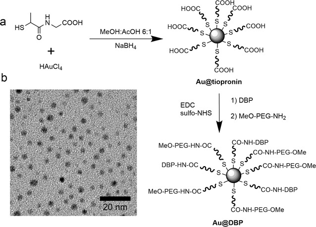
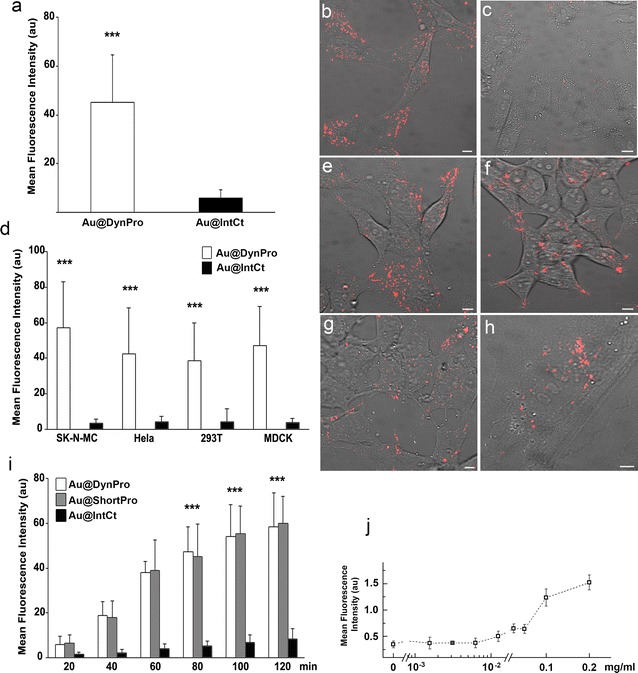
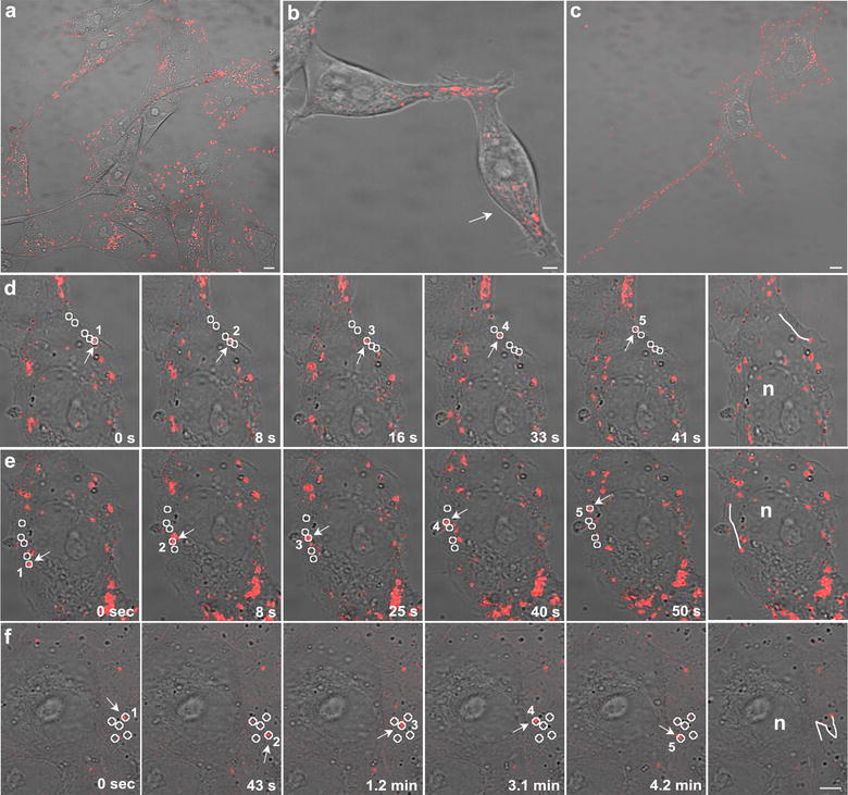
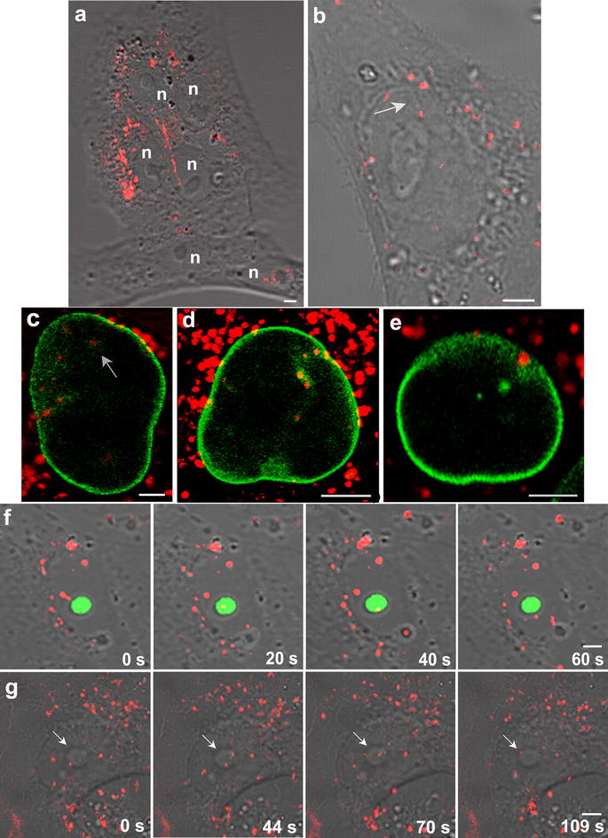
Results: Here, we show that gold nanoparticles modified with dynein-binding delivery sequences become mobile, powered by molecular motor proteins. Modified nanoparticles showed dynamic properties, such as travelling the cytosol, crossing intracellular barriers and shuttling the nuclear membrane. Furthermore, nanoparticles were transported from one cell to another through cell-to-cell contacts and quickly spread to distant cells through cell projections.
Conclusions: The capacity of these motor-bound nanoparticles to spread to many cells and increasing cellular retention, thus avoiding losses and allowing lower dosage, could make them candidate carriers for drug delivery.
Keywords: Biomimetic synthetic peptides; Drug delivery; Dynein; Microtubule motors; Nanoparticles.
Figures

Similar articles
-
In vivo characterization of dynein-driven nanovectors using Drosophila oocytes.PLoS One. 2013 Dec 12;8(12):e82908. doi: 10.1371/journal.pone.0082908. eCollection 2013. PLoS One. 2013. PMID: 24349395 Free PMC article.
-
Nano-Particles Carried by Multiple Dynein Motors Self-Regulate Their Number of Actively Participating Motors.Int J Mol Sci. 2021 Aug 18;22(16):8893. doi: 10.3390/ijms22168893. Int J Mol Sci. 2021. PMID: 34445598 Free PMC article.
-
Gold nanoparticles stabilize peptide-drug-conjugates for sustained targeted drug delivery to cancer cells.J Nanobiotechnology. 2018 Mar 30;16(1):34. doi: 10.1186/s12951-018-0362-1. J Nanobiotechnology. 2018. PMID: 29602308 Free PMC article.
-
How Dynein Moves Along Microtubules.Trends Biochem Sci. 2016 Jan;41(1):94-105. doi: 10.1016/j.tibs.2015.11.004. Epub 2015 Dec 9. Trends Biochem Sci. 2016. PMID: 26678005 Free PMC article. Review.
-
Peptide mediated formation of noble metal nanoparticles-controlling size and spatial arrangement.Curr Opin Chem Biol. 2017 Oct;40:138-144. doi: 10.1016/j.cbpa.2017.09.005. Epub 2017 Sep 27. Curr Opin Chem Biol. 2017. PMID: 28961470 Review.
Cited by
-
Viral Mimicry as a Design Template for Nucleic Acid Nanocarriers.Front Chem. 2021 Mar 10;9:613209. doi: 10.3389/fchem.2021.613209. eCollection 2021. Front Chem. 2021. PMID: 33777893 Free PMC article. Review.
-
Biomedical nanoparticle design: What we can learn from viruses.J Control Release. 2021 Jan 10;329:552-569. doi: 10.1016/j.jconrel.2020.09.045. Epub 2020 Sep 30. J Control Release. 2021. PMID: 33007365 Free PMC article. Review.
-
Effect of Clinically Used Microtubule Targeting Drugs on Viral Infection and Transport Function.Int J Mol Sci. 2022 Mar 22;23(7):3448. doi: 10.3390/ijms23073448. Int J Mol Sci. 2022. PMID: 35408808 Free PMC article.
-
The advancement of biosensor design and construction utilizing biomolecular motors.Synth Syst Biotechnol. 2025 Feb 18;10(2):543-554. doi: 10.1016/j.synbio.2025.02.007. eCollection 2025 Jun. Synth Syst Biotechnol. 2025. PMID: 40092161 Free PMC article. Review.
-
The Peptide Functionalized Inorganic Nanoparticles for Cancer-Related Bioanalytical and Biomedical Applications.Molecules. 2021 May 27;26(11):3228. doi: 10.3390/molecules26113228. Molecules. 2021. PMID: 34072160 Free PMC article. Review.
References
MeSH terms
Substances
Grants and funding
- AGL2012-34533/Ministerio de economia, industria y competitividad de España
- AGL2015-69598-R/Ministerio de economia, industria y competitividad de España
- SAF2014-54763-C2-2-R/Ministerio de economia, industria y competitividad
- 239931-NANOPUZZLE/European Research Council/International
- CA15138 Transautophagy/UE COST Action
LinkOut - more resources
Full Text Sources
Other Literature Sources
