

Lasiopodomys fuscus as an important intermediate host for Echinococcus multilocularis: isolation and phylogenetic identification of the parasite
- PMID: 29602313
- PMCID: PMC5878421
- DOI: 10.1186/s40249-018-0409-4
Lasiopodomys fuscus as an important intermediate host for Echinococcus multilocularis: isolation and phylogenetic identification of the parasite
Abstract
Background: Echinococcus multilocularis causes alveolar echinococcosis (AE) and is widely prevalent in Qinghai Province, China, where a number of different species have been identified as hosts. However, limited information is available on the Qinghai vole (Lasiopodomys fuscus), which is hyper endemic to Qinghai Province and may represent a potential intermediate host of E. multilocularis. Thus, L. fuscus could contribute to the endemicity of AE in the area.
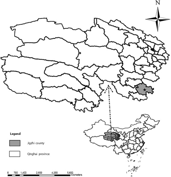
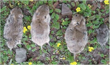
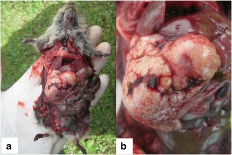
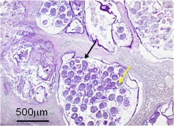
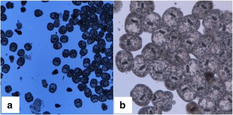
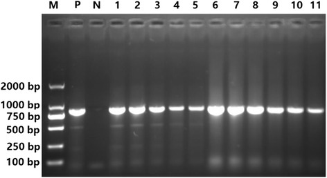
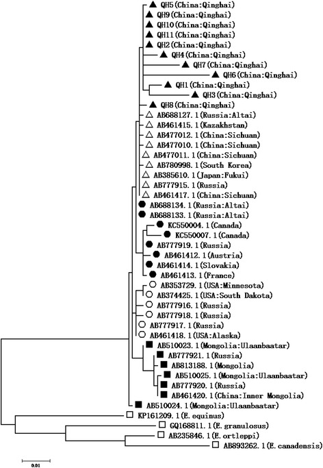
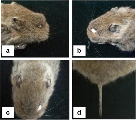
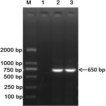
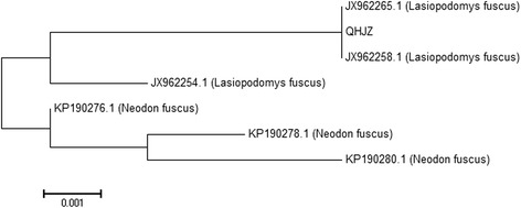
Methods: Fifty Qinghai voles were captured from Jigzhi County in Qinghai Province for the clinical identification of E. multilocularis infection via anatomical examination. Hydatid fluid was collected from vesicles of the livers in suspected voles and subjected to a microscopic examination and PCR assay based on the barcoding gene of cox 1. PCR-amplified segments were sequenced for a phylogenetic analysis. E. multilocularis-infected Qinghai voles were morphologically identified and subjected to a phylogenetic analysis to confirm their identities.
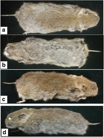
Results: Seventeen of the 50 Qinghai voles had E. multilocularis-infection-like vesicles in their livers. Eleven out of the 17 Qinghai voles presented E. multilocularis infection, which was detected by PCR and sequencing. The phylogenetic analysis showed that all 11 positive samples belonged to the E. multilocularis Asian genotype. A morphological identification and phylogenetic analysis of the E. multilocularis-infected Qinghai voles confirmed that all captured animals were L. fuscus.
Conclusions: L. fuscus can be infected with E. multilocularis and plays a potential role in the life cycle and epidemiology of E. multilocularis in the Qinghai-Tibetan Plateau of China.
Keywords: Alveolar echinococcosis; Echinococcus multilocularis; Lasiopodomys fuscus; PCR; Phylogenetic analysis; Qinghai voles; Sequencing.
Conflict of interest statement
Ethics approval and consent to participate
No specific permits were required for this study. The study did not involve endangered or protected species.
Consent for publication
Not applicable.
Competing interests
The authors declare that they have no competing interests.
Figures

Similar articles
-
Echinococcus multilocularis and Echinococcus shiquicus in a small mammal community on the eastern Tibetan Plateau: host species composition, molecular prevalence, and epidemiological implications.Parasit Vectors. 2018 May 16;11(1):302. doi: 10.1186/s13071-018-2873-x. Parasit Vectors. 2018. PMID: 29769131 Free PMC article.
-
Morphological and molecular characteristics of Echinococcus multilocularis and Echinococcus granulosus mixed infection in a dog from Xinjiang, China.Vet Parasitol. 2006 Jun 30;139(1-3):244-8. doi: 10.1016/j.vetpar.2006.03.003. Epub 2006 Apr 17. Vet Parasitol. 2006. PMID: 16616990
-
First detection of Echinococcus multilocularis in rodent intermediate hosts in Turkey.Parasitology. 2017 Nov;144(13):1821-1827. doi: 10.1017/S0031182017001226. Epub 2017 Aug 11. Parasitology. 2017. PMID: 28799893
-
Assessment of the global pattern of genetic diversity in Echinococcus multilocularis inferred by mitochondrial DNA sequences.Vet Parasitol. 2018 Oct 15;262:30-41. doi: 10.1016/j.vetpar.2018.09.013. Epub 2018 Sep 27. Vet Parasitol. 2018. PMID: 30389009 Review.
-
[Epidemiological characteristics of canine Echinococcus infection in Qinghai-Tibet Plateau of China].Zhongguo Xue Xi Chong Bing Fang Zhi Za Zhi. 2017 Apr 7;29(2):129-138. doi: 10.16250/j.32.1374.2017004. Zhongguo Xue Xi Chong Bing Fang Zhi Za Zhi. 2017. PMID: 29469312 Review. Chinese.
Cited by
-
Societal drivers of human echinococcosis in China.Parasit Vectors. 2022 Oct 22;15(1):385. doi: 10.1186/s13071-022-05480-8. Parasit Vectors. 2022. PMID: 36271415 Free PMC article.
-
Geographic distribution and prevalence of human echinococcosis at the township level in the Tibet Autonomous Region.Infect Dis Poverty. 2022 Jan 21;11(1):10. doi: 10.1186/s40249-022-00933-9. Infect Dis Poverty. 2022. PMID: 35063031 Free PMC article.
-
Species Composition of a Small Mammal Community and Prevalence of Echinococcus spp. in the Alpine Pastoral Area of the Eastern Tibetan Plateau.Pathogens. 2024 Jul 2;13(7):558. doi: 10.3390/pathogens13070558. Pathogens. 2024. PMID: 39057785 Free PMC article.
-
A new intermediate host of Echinococcus shiquicus in Qinghai-Tibet Plateau, China.Parasitol Res. 2025 Jun 18;124(6):66. doi: 10.1007/s00436-025-08494-0. Parasitol Res. 2025. PMID: 40528002 Free PMC article.
References
-
- Malikova MS, Frolova Iu V, Raskin VV, Dzemeshkevich AS, Voronina TS, Parshin VD, et al. The simultaneous surgery of heart and echinococcosis under artificial blood circulation. Khirurgiia. 2012;2012:79–82. - PubMed
MeSH terms
Substances
Supplementary concepts
Grants and funding
LinkOut - more resources
Full Text Sources
Other Literature Sources
