. 2018 May 18;293(20):7777-7785.
doi: 10.1074/jbc.RA118.002367. Epub 2018 Mar 30.

Thylakoid membranes contain a non-selective channel permeable to small organic molecules

Affiliations

Thylakoid membranes contain a non-selective channel permeable to small organic molecules

Seiji Kojima et al. J Biol Chem. .

Abstract

The thylakoid lumen is a membrane-enclosed aqueous compartment. Growing evidence indicates that the thylakoid lumen is not only a sink for protons and inorganic ions translocated during photosynthetic reactions but also a place for metabolic activities, e.g. proteolysis of photodamaged proteins, to sustain efficient photosynthesis. However, the mechanism whereby organic molecules move across the thylakoid membranes to sustain these lumenal activities is not well understood. In a recent study of Cyanophora paradoxa chloroplasts (muroplasts), we fortuitously detected a conspicuous diffusion channel activity in the thylakoid membranes. Here, using proteoliposomes reconstituted with the thylakoid membranes from muroplasts and from two other phylogenetically distinct organisms, cyanobacterium Synechocystis sp. PCC 6803 and spinach, we demonstrated the existence of nonselective channels large enough for enabling permeation of small organic compounds (e.g. carbohydrates and amino acids with Mr < 1500) in the thylakoid membranes. Moreover, we purified, identified, and characterized a muroplast channel named here CpTPOR. Osmotic swelling experiments revealed that CpTPOR forms a nonselective pore with an estimated radius of ∼1.3 nm. A lipid bilayer experiment showed variable-conductance channel activity with a typical single-channel conductance of 1.8 nS in 1 m KCl with infrequent closing transitions. The CpTPOR amino acid sequence was moderately similar to that of a voltage-dependent anion-selective channel of the mitochondrial outer membrane, although CpTPOR exhibited no obvious selectivity for anions and no voltage-dependent gating. We propose that transmembrane diffusion pathways are ubiquitous in the thylakoid membranes, presumably enabling rapid transfer of various metabolites between the lumen and stroma.

Keywords: CpTPOR; chloroplast; cyanobacteria; diffusion channel; liposome; membrane transport; muroplast; permeability; photosynthesis; porin; thylakoid membrane.


Conflict of interest statement

The authors declare that they have no conflicts of interest with the contents of this article

Figures

Figure 1.
Figure 1.
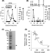
Detection of diffusion channel activity in the thylakoid membranes of muroplasts, PCC 6803, and S. oleracea chloroplasts. A, separation of the membranes of muroplasts (i), PCC 6803 (ii), and S. oleracea chloroplasts (iii) by sucrose density gradient centrifugation. IM, inner membrane; TM, thylakoid membrane; OM, outer membrane. B, diffusion channel activity in each membrane preparation. Proteins from each membrane preparation (5 μg) were reconstituted into liposomes, and membrane permeability was determined by a liposome-swelling assay with arabinose as a substrate. The data are presented as the means ± S.D. from three independent experiments (n = 9). ND, not detected. C, relative channel activity of the thylakoid membranes. Varying amounts of proteins from the thylakoid membranes of muroplasts (fraction 2), PCC 6803 (fraction 2), and S. oleracea chloroplasts (fraction 3) were reconstituted into liposomes, and membrane permeability was determined as above. The data are presented as the means ± S.D. from three independent experiments (n = 9).
Figure 2.
Figure 2.
Characterization of the channel property. A–C, proteins (10 μg) from the thylakoid membranes of muroplasts (fraction 2 in Fig. 1), PCC 6803 (fraction 2), and S. oleracea chloroplasts (fraction 3) were reconstituted into liposomes, and the influx rates of substrates were determined by liposome-swelling assay. The following substrates were used: 1, arabinose (Mr = 150.13); 2, glucose (Mr = 180.16); 3, GlcNAc (Mr = 221); 4, lactose (Mr = 342.3); 5, sucrose (Mr = 342.3); 6, maltose (Mr = 342.3); 7, raffinose (Mr = 594.53); 8, stachyose (Mr = 666.8); 9, dextran (Mr = ca. 1500); 10, l-Pro (Mr = 115.13); 11, l-Val (Mr = 117.15); 12, l-Asn (Mr = 132.12); 13, d-alanyl-d-alanine (Mr = 160); and 14, l-Phe (Mr = 165.19). The data are presented as the means from two independent experiments (n = 6). The theoretical relative influx rates are shown as broken lines in each graph. The expected pore radii were 1.3 nm, 3.0 nm, and 1.7 nm for the muroplasts, PCC 6803, and S. oleracea chloroplasts, respectively. D, the relative influx rates of glucose and its anionic and di-anionic derivatives gluconate and glucarate through the thylakoid membrane channels of muroplasts, PCC 6803, and S. oleracea chloroplasts. Proteoliposomes reconstituted with each thylakoid membrane were prepared as above, and the influx rates were determined by liposome-swelling assay. The data are presented as the means ± S.D. from three independent experiments (n = 9).
Figure 3.
Figure 3.
Purification of CpTPOR. A, gel filtration chromatography of solubilized muroplast thylakoid membrane proteins. The molecular mass standards are as follows: thyroglobulin, 669 kDa; apoferritin, 443 kDa; α-amylase, 200 kDa; alcohol dehydrogenase, 150 kDa; albumin, 66 kDa; and carbonic anhydrase, 29 kDa. The channel activity of each fraction was examined by liposome-swelling assay with arabinose as a substrate. B, anion exchange chromatography of the 23- to 25-min elution fraction obtained after gel filtration chromatography. The timetable of the LiCl gradient is shown above the graph. The channel activity of each fraction was examined as described above. C, SDS-PAGE analysis of the purified CpTPOR preparation. The gel was stained with Coomassie Brilliant Blue. M, molecular mass standards. D, the channel property of the purified CpTPOR. The purified CpTPOR protein (3 μg) was reconstituted into liposomes, and the influx rates of the substrates were determined by liposome-swelling assay. The following substrates were used: 1, arabinose (Mr = 150.13); 2, glucose (Mr = 180.16); 3, GlcNAc (Mr = 221); 4, lactose (Mr = 342.3); 5, sucrose (Mr = 342.3); 6, maltose (Mr = 342.3); 7, raffinose (Mr = 594.53); 8, stachyose (Mr = 666.8); 9, dextran (Mr = approximately 1500); 10, l-Pro (Mr = 115.13); 11, l-Val (Mr = 117.15); 12, l-Asn (Mr = 132.12); 13, d-alanyl-d-alanine (Mr = 160); and 14, l-Phe (Mr = 165.19). The data are presented as the means from triplicate measurements (n = 3).The theoretical relative influx rate assuming a pore radius of 1.3 nm is shown by a broken line.
Figure 4.
Figure 4.
The CpTPOR sequence characteristics. The predicted secondary structure is shown underneath the amino acid sequence. The putative α-helix is shown as a gray box, and the putative β-strands are shown as arrows. The putative VDAC motifs, listed in Table S2, are boxed.
Figure 5.
Figure 5.
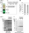
The evaluation of mitochondrial contamination in the muroplast preparation. A, muroplasts were isolated from C. paradoxa cells and purified by Percoll gradient centrifugation (i), and the cytochrome c oxidase activity in each fraction was determined (ii). The data are presented as the means ± S.D. from three independent experiments (n = 9). ND, not detected. B, diffusion channel activities of the thylakoid membranes of muroplasts collected from fractions B and C after Percoll gradient centrifugation. The proteins of each thylakoid membrane (5 μg) were reconstituted into liposomes, and the diffusion channel activity was determined by liposome-swelling assay with arabinose as a substrate. The data are presented as the means ± S.D. from triplicate determinations (n = 3). C, detection of CpTPOR by immunoblotting. Fifteen micrograms of proteins of the total membranes in fraction A (lane I) and the thylakoid membranes isolated from the muroplasts in fractions B and C (lanes II and III, respectively) were separated by SDS-PAGE (i). Proteins were then blotted onto a polyvinylidene fluoride membrane, and CpTPOR was detected using anti-CpTPOR antibody and alkaline phosphatase-conjugated anti-rabbit IgG antibody (ii). M, molecular mass standards; CBB, Coomassie Brilliant Blue.
Figure 6.
Figure 6.
The electrophysiological characterization of CpTPOR. A, typical current traces of CpTPOR. The single-channel conductance of purified CpTPOR was determined in 1 m KCl at various membrane voltages using the contact bubble bilayer method. B, the current-voltage relationship of CpTPOR determined in 1 m KCl. The data are presented as the current amplitude of the predominant conductance level for each membrane potential. C, representative subconductance current traces of CpTPOR, observed in 1 m KCl with +100 mV membrane potential. D, typical current trace of CpTPOR observed in 50 mm KCl with +50 mV membrane potential.

Similar articles

Cited by

References

    1. Engel B. D., Schaffer M., Kuhn Cuellar L., Villa E., Plitzko J. M., and Baumeister W. (2015) Native architecture of the Chlamydomonas chloroplast revealed by in situ cryo-electron tomography. eLife 4, e04889 10.7554/eLife.04889 - DOI - PMC - PubMed
    1. Nevo R., Charuvi D., Shimoni E., Schwarz R., Kaplan A., Ohad I., and Reich Z. (2007) Thylakoid membrane perforations and connectivity enable intracellular traffic in cyanobacteria. EMBO J. 26, 1467–1473 10.1038/sj.emboj.7601594 - DOI - PMC - PubMed
    1. Ting C. S., Hsieh C., Sundararaman S., Mannella C., and Marko M. (2007) Cryo-electron tomography reveals the comparative three-dimensional architecture of Prochlorococcus, a globally important marine cyanobacterium. J. Bacteriol. 189, 4485–4493 10.1128/JB.01948-06 - DOI - PMC - PubMed
    1. Konorty M., Kahana N., Linaroudis A., Minsky A., and Medalia O. (2008) Structural analysis of photosynthetic membranes by cryo-electron tomography of intact Rhodopseudomonas viridis cells. J. Struct. Biol. 161, 393–400 10.1016/j.jsb.2007.09.014 - DOI - PubMed
    1. Järvi S., Gollan P. J., and Aro E. M. (2013) Understanding the roles of the thylakoid lumen in photosynthesis regulation. Front. Plant Sci. 4, 434 - PMC - PubMed

Publication types

LinkOut - more resources