

Exploring the role of post-translational modifications in regulating α-synuclein interactions by studying the effects of phosphorylation on nanobody binding
- PMID: 29603451
- PMCID: PMC6032363
- DOI: 10.1002/pro.3412
Exploring the role of post-translational modifications in regulating α-synuclein interactions by studying the effects of phosphorylation on nanobody binding
Abstract
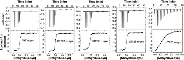
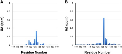
Intracellular deposits of α-synuclein in the form of Lewy bodies are major hallmarks of Parkinson's disease (PD) and a range of related neurodegenerative disorders. Post-translational modifications (PTMs) of α-synuclein are increasingly thought to be major modulators of its structure, function, degradation and toxicity. Among these PTMs, phosphorylation near the C-terminus at S129 has emerged as a dominant pathogenic modification as it is consistently observed to occur within the brain and cerebrospinal fluid (CSF) of post-mortem PD patients, and its level appears to correlate with disease progression. Phosphorylation at the neighboring tyrosine residue Y125 has also been shown to protect against α-synuclein toxicity in a Drosophila model of PD. In the present study we address the potential roles of C-terminal phosphorylation in modulating the interaction of α-synuclein with other protein partners, using a single domain antibody fragment (NbSyn87) that binds to the C-terminal region of α-synuclein with nanomolar affinity. The results reveal that phosphorylation at S129 has negligible effect on the binding affinity of NbSyn87 to α-synuclein while phosphorylation at Y125, only four residues away, decreases the binding affinity by a factor of 400. These findings show that, despite the fact that α-synuclein is intrinsically disordered in solution, selective phosphorylation can modulate significantly its interactions with other molecules and suggest how this particular form of modification could play a key role in regulating the normal and aberrant function of α-synuclein.
Keywords: Parkinson's disease; isothermal titration calorimetry; nanobody); nuclear magnetic resonance; phosphorylation; protein misfolding; single-domain antibody (sdAb; surface plasmon resonance; α-synuclein.
© 2018 The Protein Society.
Figures


Similar articles
-
Phosphorylation of α-Synuclein at Y125 and S129 alters its metal binding properties: implications for understanding the role of α-Synuclein in the pathogenesis of Parkinson's Disease and related disorders.ACS Chem Neurosci. 2011 Nov 16;2(11):667-75. doi: 10.1021/cn200074d. Epub 2011 Sep 14. ACS Chem Neurosci. 2011. PMID: 22860160 Free PMC article.
-
C-Terminal Tyrosine Residue Modifications Modulate the Protective Phosphorylation of Serine 129 of α-Synuclein in a Yeast Model of Parkinson's Disease.PLoS Genet. 2016 Jun 24;12(6):e1006098. doi: 10.1371/journal.pgen.1006098. eCollection 2016 Jun. PLoS Genet. 2016. PMID: 27341336 Free PMC article.
-
Investigating the presence of doubly phosphorylated α-synuclein at tyrosine 125 and serine 129 in idiopathic Lewy body diseases.Brain Pathol. 2020 Jul;30(4):831-843. doi: 10.1111/bpa.12845. Epub 2020 May 6. Brain Pathol. 2020. PMID: 32324926 Free PMC article.
-
Effects of alpha-synuclein post-translational modifications on metal binding.J Neurochem. 2019 Sep;150(5):507-521. doi: 10.1111/jnc.14721. Epub 2019 Jul 9. J Neurochem. 2019. PMID: 31099098 Review.
-
The role of posttranslational modifications of α-synuclein and LRRK2 in Parkinson's disease: Potential contributions of environmental factors.Biochim Biophys Acta Mol Basis Dis. 2019 Aug 1;1865(8):1992-2000. doi: 10.1016/j.bbadis.2018.11.017. Epub 2018 Nov 24. Biochim Biophys Acta Mol Basis Dis. 2019. PMID: 30481588 Free PMC article. Review.
Cited by
-
Characterizing the inhibition of α-synuclein oligomerization by a pharmacological chaperone that prevents prion formation by the protein PrP.Protein Sci. 2019 Sep;28(9):1690-1702. doi: 10.1002/pro.3684. Epub 2019 Aug 2. Protein Sci. 2019. PMID: 31306510 Free PMC article.
-
Nanobodies Right in the Middle: Intrabodies as Toolbox to Visualize and Modulate Antigens in the Living Cell.Biomolecules. 2020 Dec 21;10(12):1701. doi: 10.3390/biom10121701. Biomolecules. 2020. PMID: 33371447 Free PMC article. Review.
-
Phosphatidylinositol-3,4,5-trisphosphate interacts with alpha-synuclein and initiates its aggregation and formation of Parkinson's disease-related fibril polymorphism.Acta Neuropathol. 2023 May;145(5):573-595. doi: 10.1007/s00401-023-02555-3. Epub 2023 Mar 20. Acta Neuropathol. 2023. PMID: 36939875 Free PMC article.
-
Specific Detection of Physiological S129 Phosphorylated α-Synuclein in Tissue Using Proximity Ligation Assay.J Parkinsons Dis. 2023;13(2):255-270. doi: 10.3233/JPD-213085. J Parkinsons Dis. 2023. PMID: 36847016 Free PMC article.
-
In Search of Effective Treatments Targeting α-Synuclein Toxicity in Synucleinopathies: Pros and Cons.Front Cell Dev Biol. 2020 Sep 4;8:559791. doi: 10.3389/fcell.2020.559791. eCollection 2020. Front Cell Dev Biol. 2020. PMID: 33015057 Free PMC article. Review.
References
-
- Kahle PJ, Neumann M, Ozmen L, Muller V, Jacobsen H, Schindzielorz A, Okochi M, Leimer U, van Der Putten H, Probst A, Kremmer E, Kretzschmar HA, Haass C (2000) Subcellular localization of wild‐type and Parkinson's disease‐associated mutant alpha‐synuclein in human and transgenic mouse brain. J Neurosci 20:6365–6373. - PMC - PubMed
-
- Galvin JE, Lee VM, Trojanowski JQ (2001) Synucleinopathies: clinical and pathological implications. Arch Neurol 58:186–190. - PubMed
-
- Martí MJ, Tolosa E, Campdelacreu J (2003) Clinical overview of the synucleinopathies. Mov Disord 18:21–27. - PubMed
-
- Wakabayashi K, Tanji K, Mori F, Takahashi H (2007) The Lewy body in Parkinson's disease: molecules implicated in the formation and degradation of alpha‐synuclein aggregates. Neuropathology 27:494–506. - PubMed
Publication types
MeSH terms
Substances
Grants and funding
LinkOut - more resources
Full Text Sources
Other Literature Sources
Miscellaneous
