. 2018 Jun;17(3):e12758.
doi: 10.1111/acel.12758. Epub 2018 Mar 30.

Involvement of serum-derived exosomes of elderly patients with bone loss in failure of bone remodeling via alteration of exosomal bone-related proteins

Affiliations

Involvement of serum-derived exosomes of elderly patients with bone loss in failure of bone remodeling via alteration of exosomal bone-related proteins

Yong Xie et al. Aging Cell. 2018 Jun.

Abstract

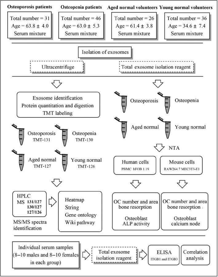
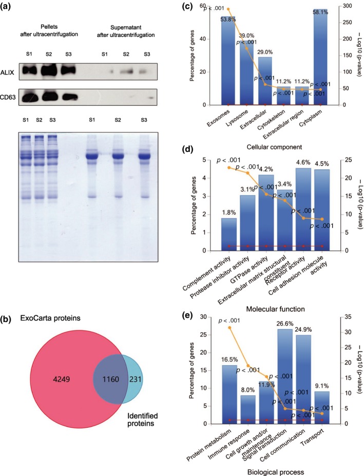
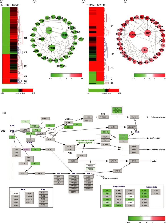
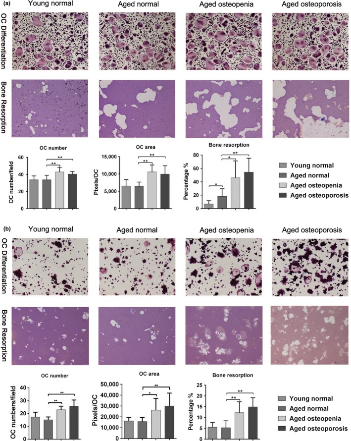
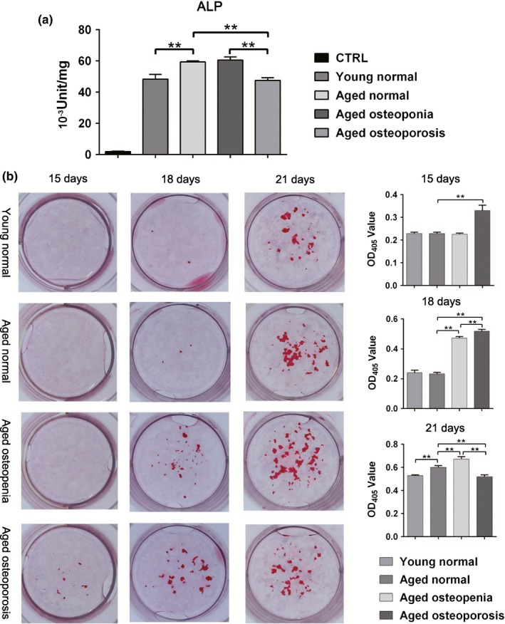
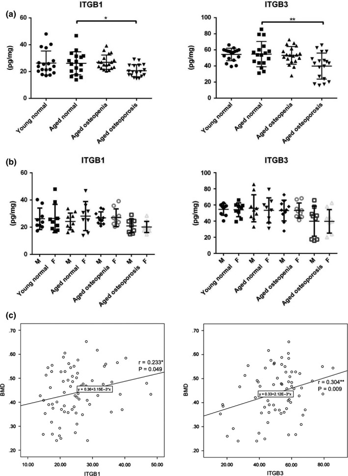
Exosomes are secreted into the blood by various types of cells. These extracellular vesicles are involved in the contribution of exosomal proteins to osteoblastic or osteoclastic regulatory networks during the failure of bone remodeling, which results in age-related bone loss. However, the molecular changes in serum-derived exosomes (SDEs) from aged patients with low bone density and their functions in bone remodeling remain to be fully elucidated. We present a quantitative proteomics analysis of exosomes purified from the serum of the elderly patients with osteoporosis/osteopenia and normal volunteers; these data are available via Proteome Xchange with the identifier PXD006463. Overall, 1,371 proteins were identified with an overlap of 1,160 Gene IDs among the ExoCarta proteins. Bioinformatics analysis and in vitro studies suggested that protein changes in SDEs of osteoporosis patients are not only involved in suppressing the integrin-mediated mechanosensation and activation of osteoblastic cells, but also trigger the differentiation and resorption of osteoclasts. In contrast, the main changes in SDEs of osteopenia patients facilitated both activation of osteoclasts and formation of new bone mass, which could result in a compensatory elevation in bone remodeling. While the SDEs from aged normal volunteers might play a protective role in bone health through facilitating adhesion of bone cells and suppressing aging-associated oxidative stress. This information will be helpful in elucidating the pathophysiological functions of SDEs and aid in the development of senile osteoporosis diagnostics and therapeutics.

Keywords: bone remodeling; exosome; osteoblasts; osteoclasts; osteoporosis.


Figures

Figure 1
Figure 1
Experimental workflow. “OC” refers to osteoclast, “NTA” refers to nanoparticle tracking analysis
Figure 2
Figure 2
Efficiency of SDE extraction and quantitative characteristics of protein profiles. All identified proteins have been submitted to the ExoCarta database and GO classification system. (a) Western blots showing enrichment of the exosome marker CD63 and ALIX in exosome pellets but not in the supernatants of pooled serum samples after ultracentrifugation. Coomassie brilliant blue staining was used as a control to assess standardized loading. (b) Venn diagram showing the overlap of identified proteins with ExoCarta proteins (5,409; Release date: 29 July 2015). (c–e) The six most enriched categories and the enrichment significance (−log (p‐value), < .05) of identified proteins in cellular components (c), molecular functions (d), and biological process (e) categories. The percentage of proteins identified in each category is indicated. “S1” refers to pooled serum from Aged normal volunteers, “S2” refers to pooled serum from Aged osteopenia patients, “S3” refers to pooled serum from Aged osteoporosis patients
Figure 3
Figure 3
Bioinformatics analysis of DEPs from SDEs based on STRING and Wiki pathway databases. The heatmap was constructed into clusters by Hierarchical Clustering Explorer 3.5 and shows contrasting or similar expression levels of osteoporosis‐associated SDE DEPs compared with those identified from SDEs of osteopenia patients (MS data presented as the ratios to 131/127 and 130/127 were input into Hierarchical Clustering Explorer 3.5 with Ln transformation). The DEPs in the protein–protein interaction networks are shown as nodes (MS data presented as the ratios to 131/127 and 130/127 were matched to STRING networks with log2 transformation). Up‐ or downregulation of identified proteins is indicated by colors either in the heatmap or networks (upregulated in red, downregulated in green). All identified proteins were mapped to the relevant Wiki pathway database. Proteins are represented by boxes labeled with the protein name. Relative protein expression level in SDEs of osteoporosis patients is indicated by colors (MS data presented as the ratios to 131/127 and 130/127 were matched to STRING networks with log2 transformation). Proteins in gray were not identified in this study. (a) Heatmap of DEPs of SDEs from patients with osteoporosis (131/127) comparing to those from patients with osteopenia (130/127). (b) Network of overall DEPs in SDEs of patients with osteoporosis. (c) Heatmap of DEPs of SDEs from patients with osteopenia (130/127) comparing to those from patients with osteopenia (131/127). (d) Network of upregulated proteins in SDEs of patients with osteopenia. (e) DEPs of SDEs from osteoporosis patients mapping to the Integrin‐mediated cell adhesion pathway. “C1‐6” refers to cluster 1‐6 in each heatmap
Figure 4
Figure 4
SDEs regulate osteoclast differentiation and bone resorption. (a) The number and area of rearrangement of TRAP‐positive multinucleated osteoclasts cultured with SDEs from osteoporosis or osteopenia patients were significantly higher than that of TRAP‐positive multinucleated osteoclasts cultured with SDEs from aged normal volunteers. Moreover, the bone resorption of osteoclasts cultured with SDEs from osteoporosis or osteopenia patients was significantly greater than the resorption of osteoclasts cultured with aged normal volunteers. In addition, the bone resorption of osteoclasts cultured with SDEs from aged normal volunteers was significantly greater than the resorption of osteoclasts cultured with SDEs from young normal volunteers. However, there was no significant difference in the number and area of rearrangement of between the TRAP‐positive osteoclasts treated with either SDEs from young or aged normal volunteers. (b) RAW264.7 cells cultured with RANKL had a similar effect on osteoclast differentiation and bone resorption compared with the effects of SDEs from different groups; however, the bone resorption of osteoclasts cultured with SDEs from aged normal volunteers was no difference comparing to the resorption of osteoclasts cultured with SDEs from young normal volunteers. Representative photographs are shown in the up panel. Quantification of cells is shown in the down panel. All values are representative of at least two independent experiments with similar results and are displayed as mean ± SD. **p < .01, *p < .05. “OC” refers to osteoclast
Figure 5
Figure 5
SDEs regulate bone formation by osteoblasts. (a) In the human osteoblastic cell line Hob (hFOB 1.19) cultured on day 21, the alkaline phosphatase (ALP) levels confirmed that higher levels of mineralization of cells cultured with SDEs from osteopenia patients than that of cells cultured with SDEs from aged normal volunteers. However, the level of mineralization in hFOB 1.19 cells cultured with SDEs from osteoporosis patients was significantly lower than that of cells cultured with SDEs from aged normal volunteers. In addition, the mineralization level of cells cultured with SDEs from aged normal was higher than that of cells cultured with SDEs from young normal volunteers. (b) At 15 days after induction of MC3T3‐E1 cells by osteogenic media containing vitamin C and β‐glycerol phosphate, mineralization nodules were first observed in the group cultured in the presence of SDEs from osteoporosis patients. Three days later, mineralization nodules were observed in all the groups, and the OD values of Alizarin Red staining in the Aged osteoporosis and osteopenia groups were higher than those of the Aged normal group. In the later stages of matrix mineralization on day 21, the OD value in the Aged osteoporosis group was lower than those in both the Aged normal and osteopenia groups. Moreover, the OD value in the Aged osteopenia group was higher than that in the Aged normal group. The OD value in the Young normal group was lower than that in the Aged normal group. The OD values of Alizarin Red staining in all these four groups were in accordance with the ALP levels detected in human osteoblastic cells. Representative images are shown in the left panel. Quantification of cells is shown in the right panel. All values are representative of at least two independent experiments with similar results and are displayed as mean ± SD. **p < .01, *p < .05
Figure 6
Figure 6
Verification of protein expression levels by ELISA and correlation analysis between the changes in exosomal proteins and BMD. To validate the MS results, (a) the SDEs of 72 serum samples were subjected to ELISA using antibodies for specific detection of integrin β1 (ITGB1) and integrin β3 (ITGB3), individually. The fluctuation in the levels of these proteins was consistent with the proteomics data. (b) The data also demonstrated that there was no significant difference between the male and female patients in each group. (c) Analysis of the correlation between the expression level of ITGB1 and ITGB3 in SDEs and BMD. “BMD” refers to bone mass density

Similar articles

Cited by

References

    1. Barker, A. L. , McNeil, J. J. , Seeman, E. , Ward, S. A. , Sanders, K. M. , Khosla, S. , … Talevski, J. (2016). A randomised controlled trial of low‐dose aspirin for the prevention of fractures in healthy older people: Protocol for the ASPREE‐Fracture substudy. Injury Prevention: Journal of the International Society for Child and Adolescent Injury Prevention, 22, 297–301. 10.1136/injuryprev-2015-041655 - DOI - PMC - PubMed
    1. Beukhof, C. M. , Medici, M. , van den Beld, A. W. , Hollenbach, B. , Hoeg, A. , Visser, W. E. , … Peeters, R. P. (2016). Selenium status is positively associated with bone mineral density in healthy aging European men. PLoS ONE, 11, e0152748 10.1371/journal.pone.0152748 - DOI - PMC - PubMed
    1. Cao, J. J. , Gregoire, B. R. , & Zeng, H. (2012). Selenium deficiency decreases antioxidative capacity and is detrimental to bone microarchitecture in mice. The Journal of Nutrition, 142, 1526–1531. 10.3945/jn.111.157040 - DOI - PubMed
    1. Chen, Y. , Xie, Y. , Xu, L. , Zhan, S. , Xiao, Y. , Gao, Y. , … Ge, W. (2017). Protein content and functional characteristics of serum‐purified exosomes from patients with colorectal cancer revealed by quantitative proteomics. International Journal of Cancer, 140, 900–913. 10.1002/ijc.30496 - DOI - PubMed
    1. Cheung, W. Y. , Simmons, C. A. , & You, L. (2012). Osteocyte apoptosis regulates osteoclast precursor adhesion via osteocytic IL‐6 secretion and endothelial ICAM‐1 expression. Bone, 50, 104–110. 10.1016/j.bone.2011.09.052 - DOI - PubMed

Publication types