. 2018 Dec;596(23):6105-6119.
doi: 10.1113/JP275659. Epub 2018 Apr 29.

Altered autonomic control of heart rate variability in the chronically hypoxic fetus

Affiliations

Altered autonomic control of heart rate variability in the chronically hypoxic fetus

C J Shaw et al. J Physiol. 2018 Dec.

Abstract

Key points: Fetal heart rate variability (FHRV) has long been recognised as a powerful predictor of fetal wellbeing, and a decrease in FHRV is associated with fetal compromise. However, the mechanisms by which FHRV is reduced in the chronically hypoxic fetus have yet to be established. The sympathetic and parasympathetic influences on heart rate mature at different rates throughout fetal life, and can be assessed by time domain and power spectral analysis of FHRV. In this study of chronically instrumented fetal sheep in late gestation, we analysed FHRV daily over a 16 day period towards term, and compared changes between fetuses of control and chronically hypoxic pregnancy. We show that FHRV in sheep is reduced by chronic hypoxia, predominantly due to dysregulation of the sympathetic control of the fetal heart rate. This presents a potential mechanism by which a reduction in indices of FHRV predicts fetuses at increased risk of neonatal morbidity and mortality in humans. Reduction in overall FHRV may therefore provide a biomarker that autonomic dysregulation of fetal heart rate control has taken place in a fetus where uteroplacental dysfunction is suspected.

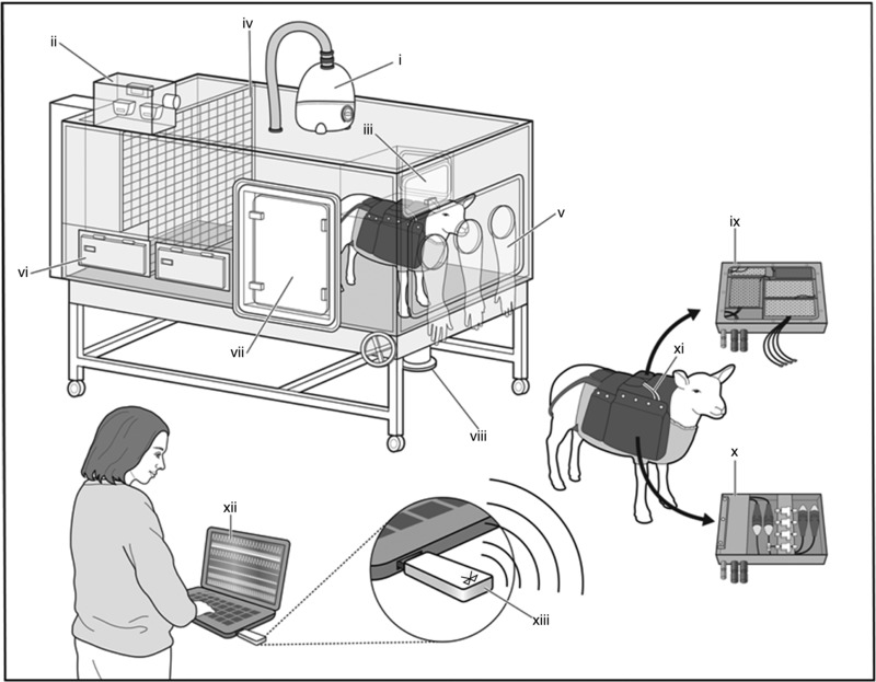
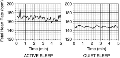
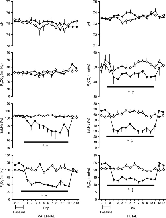
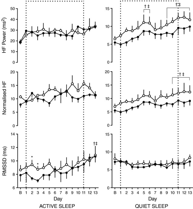
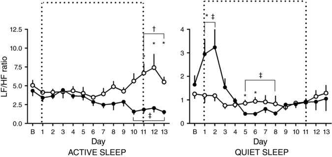
Abstract: Although fetal heart rate variability (FHRV) has long been recognised as a powerful predictor of fetal wellbeing, the mechanisms by which it is reduced in the chronically hypoxic fetus have yet to be established. In particular, the physiological mechanism underlying the reduction of short term variation (STV) in fetal compromise remains unclear. In this study, we present a longitudinal study of the development of autonomic control of FHRV, assessed by indirect indices, time domain and power spectral analysis, in normoxic and chronically hypoxic, chronically catheterised, singleton fetal sheep over the last third of gestation. We used isobaric chambers able to maintain pregnant sheep for prolonged periods in hypoxic conditions (stable fetal femoral arterial PO2 10-12 mmHg), and a customised wireless data acquisition system to record beat-to-beat variation in the fetal heart rate. We determined in vivo longitudinal changes in overall FHRV and the sympathetic and parasympathetic contribution to FHRV in hypoxic (n = 6) and normoxic (n = 6) ovine fetuses with advancing gestational age. Normoxic fetuses show gestational age-related increases in overall indices of FHRV, and in the sympathetic nervous system contribution to FHRV (P < 0.001). Conversely, gestational age-related increases in overall FHRV were impaired by exposure to chronic hypoxia, and there was evidence of suppression of the sympathetic nervous system control of FHRV after 72 h of exposure to hypoxia (P < 0.001). This demonstrates that exposure to late gestation isolated chronic fetal hypoxia has the potential to alter the development of the autonomic nervous system control of FHRV in sheep. This presents a potential mechanism by which a reduction in indices of FHRV in human fetuses affected by uteroplacental dysfunction can predict fetuses at increased risk.

Keywords: Fetal Growth Restriction; Fetal heart rate; Fetal heart rate variability; Intrauterine hypoxia; Sympathetic nervous system.


Figures

Figure 1
Figure 1. Isobaric hypoxic chambers and the CamDAS system
Each chamber was equipped with an electronic servo‐controlled humidity cool steam injection system to return the appropriate humidity to the inspirate (i). Ambient PO2, PCO2, humidity and temperature within each chamber were monitored via sensors (ii). For experimental procedures, each chamber had a double transfer port (iii) to internalise material and a manually operated sliding panel (iv) to bring the ewe into a position where daily sampling of blood could be achieved through glove compartments (v). Each chamber incorporated a drinking bowl on continuous water supply and a rotating food compartment (vi) for determining food intake. A sealed transfer isolation cart could be attached to a side exit (vii) to couple chambers together for cleaning. The CamDAS system was contained in a custom‐made sheep jacket able to hold the data acquisition system box (ix) in one side pouch and a box containing the pressure connectors (x) in the other. Cables (xi) connected the two boxes together and also to two battery packs able to power the system for 24 h. Measurements made using the data acquisition were transmitted wirelessly via Bluetooth (xiii) to a laptop kept outside the chamber room (xii) on which it was possible to view continuous recordings of the maternal and fetal cardiovascular data (reproduced with permission, Allison et al. 2016).
Figure 2
Figure 2. Fetal heart rate variability patterns
A representative example of active sleep (left) and quiet sleep (right) categorised by visual identification.
Figure 3
Figure 3. Maternal and fetal blood gas, acid–base status during chronic normoxia and hypoxia
Values represent the mean ± SEM (n = 12) for arterial pH, partial pressure of arterial carbon dioxide (P aC O2) and oxygen (PaO2) and percentage oxygen saturation of haemoglobin (Sat.Hb). Chronic hypoxia, filled circles; chronic normoxia, open circles. Significant differences (P < 0.05): *significant main effect of hypoxia compared with normoxia; significant main effect of time in hypoxic pregnancy on each individual day 1–13 when compared to the baseline period (RM two‐way ANOVA with post hoc Holm–Sidak and Tukey's tests).
Figure 4
Figure 4. Ontogenic changes in fetal heart rate and variability during chronic normoxia and hypoxia
Values represent the mean ± SEM (n = 12) for absolute fetal heart rate, SDNN, short term variation (STV) and total power in quiet and active sleep. Period of chronic hypoxia (filled circles) or normoxia (open circles) indicated by dashed box. ‘B’ represents an average of values taken in the 72 h period −2 to 0. Significant differences (P < 0.05): *significant effect of oxygenation between treatment groups; significant effect of gestational age in normoxic pregnancy on each individual day 1–13 when compared to the baseline ‘B’ period; significant effect of gestational age in hypoxic pregnancy on each individual day 1–13 when compared to the baseline ‘B’ period (RM two‐way ANOVA with post hoc Tukey's and Holm–Sidak tests).
Figure 5
Figure 5. Ontogenic changes in indices of sympathetic contribution to fetal heart rate variability during chronic normoxia and hypoxia
Values represent the mean ± SEM (n = 12) for absolute and normalised LF in quiet and active sleep. Period of chronic hypoxia (filled circles) or normoxia (open circles) indicated by dashed box. ‘B’ represents an average of values taken in the 72 h period −2 to 0. Significant differences (P < 0.05): *significant effect of oxygenation between treatment groups; significant effect of gestational age in normoxic pregnancy on each individual day 1–13 when compared to the baseline ‘B’ period; significant effect of gestational age in hypoxic pregnancy on each individual day 1–13 when compared to the baseline ‘B’ period (RM two‐way ANOVA with post hoc Tukey's and Holm–Sidak tests).
Figure 6
Figure 6. Ontogenic changes in indices of parasympathetic contribution to fetal heart rate variability during chronic normoxia and hypoxia
Values represent the mean ± SEM (n = 12) for RMSSD, absolute and normalised HF in quiet and active sleep. Period of chronic hypoxia (filled circles) or normoxia (open circles) indicated by dashed box. ‘B’ represents an average of values taken in the 72 h period −2 to 0. Significant differences (P < 0.05): *significant effect of oxygenation between treatment groups; significant effect of gestational age in normoxic pregnancy on each individual day 1–13 when compared to the baseline ‘B’ period; significant effect of gestational age in hypoxic pregnancy on each individual day 1–13 when compared to the baseline ‘B’ period (RM two‐way ANOVA with post hoc Tukey's and Holm–Sidak tests).
Figure 7
Figure 7. Ontogenic changes in sympathovagal balance during chronic normoxia and hypoxia
Values represent the mean ± SEM (n = 12) for the LF to HF ratio in quiet and active sleep. Period of chronic hypoxia (filled circles) or normoxia (open circles) indicated by dashed box. ‘B’ represents an average of values taken in the 72 h period −2 to 0. Significant differences (P < 0.05): *significant effect of oxygenation between treatment groups; significant effect of gestational age in normoxic pregnancy on each individual day 1–13 when compared to the baseline ‘B’ period; significant effect of gestational age in hypoxic pregnancy on each individual day 1–13 when compared to the baseline ‘B’ period (RM two‐way ANOVA with post hoc Tukey's and Holm–Sidak tests).

Comment in

Similar articles

Cited by

References

    1. Allison BJ, Brain KL, Niu Y, Kane AD, Herrera EA, Thakor AS, Botting KJ, Cross CM, Itani N, Skeffington KL, Beck C & Giussani DA (2016). Fetal in vivo continuous cardiovascular function during chronic hypoxia. J Physiol 594, 1247–1264. - PMC - PubMed
    1. Assali NS, Brinkman CR 3rd, Woods JR Jr, Dandavino A & Nuwayhid B (1977). Development of neurohumoral control of fetal, neonatal, and adult cardiovascular functions. Am J Obstet Gynecol 129, 748–759. - PubMed
    1. Bennet L & Gunn AJ (2009). The fetal heart rate response to hypoxia: insights from animal models. Clin Perinatol 36, 655–672. - PubMed
    1. Brain KL, Allison BJ, Niu Y, Cross CM, Itani N, Kane AD, Herrera EA & Giussani DA (2015). Induction of controlled hypoxic pregnancy in large mammalian species. Physiol Rep 3, e12614. - PMC - PubMed
    1. Ferrario M, Signorini MG & Magenes G (2009). Complexity analysis of the fetal heart rate variability: early identification of severe intrauterine growth‐restricted fetuses. Med Biol Eng Comput 47, 911–919. - PMC - PubMed

Publication types