

Computer tomographic imaging in 4 dogs with primary nasal canine transmissible venereal tumor and differing cellular phenotype
- PMID: 29604101
- PMCID: PMC5980456
- DOI: 10.1111/jvim.15125
Computer tomographic imaging in 4 dogs with primary nasal canine transmissible venereal tumor and differing cellular phenotype
Abstract
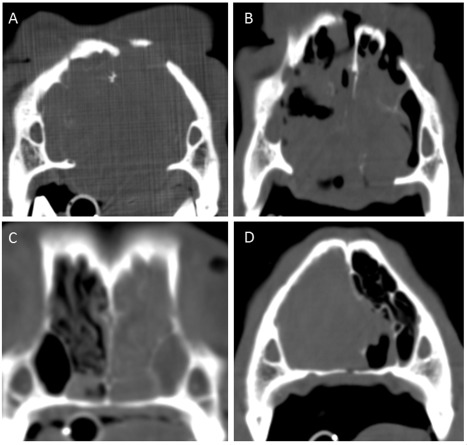
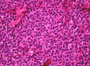
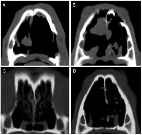
Primary nasal canine transmissible venereal tumor (CTVT) without genital affection is uncommon. The aim of this report was to describe the primary nasal CTVT findings and CT staging in 4 dogs with different cytological phenotypes. Three male dogs and 1 bitch were evaluated for their chronic histories of sneezing, snoring, mucopurulent nasal discharge and nasal deformation. Cytological examination of nasal secretions suggested CTVT, confirmed by histopathological examination and LINE-1/c-myc. Males had the plasmacytoid phenotype of CTVT, and the bitch had the lymphocytoid phenotype. CTVT were staged based on the CT findings using modified Adams staging system. The bitch was classified as stage 1, 2 males were classified as stage 3 and 1 male as stage 4. All dogs had a complete tumoral remission after chemotherapy. Plasmacytoid phenotype was identified in cases with most important damage of the nasal cavity. However, the cytological type did not affect the response to chemotherapy.
Keywords: CTVT; cytology; dogs; nasal tumor.
Copyright © 2018 The Authors. Journal of Veterinary Internal Medicine published by Wiley Periodicals, Inc. on behalf of the American College of Veterinary Internal Medicine.
Figures

Similar articles
-
Pre- and post-treatment computed tomographic findings of a primary intranasal transmissible venereal tumor in a canine patient.J Biol Regul Homeost Agents. 2018 May-Jun;32(3):571-576. J Biol Regul Homeost Agents. 2018. PMID: 29921382
-
Cell-based polymerase chain reaction for canine transmissible venereal tumor (CTVT) diagnosis.J Vet Med Sci. 2016 Aug 1;78(7):1167-73. doi: 10.1292/jvms.15-0710. Epub 2016 Apr 14. J Vet Med Sci. 2016. PMID: 27075116 Free PMC article.
-
Cell-mediated immunity and expression of MHC class I and class II molecules in dogs naturally infected by canine transmissible venereal tumor: Is there complete spontaneous regression outside the experimental CTVT?Res Vet Sci. 2022 Jul;145:193-204. doi: 10.1016/j.rvsc.2022.02.020. Epub 2022 Feb 23. Res Vet Sci. 2022. PMID: 35240476
-
Canine transmissible venereal tumor - From general to molecular characteristics: A review.Anim Genet. 2023 Feb;54(1):82-89. doi: 10.1111/age.13260. Epub 2022 Oct 19. Anim Genet. 2023. PMID: 36259378 Review.
-
Epidemiological study of canine transmissible venereal tumor (CTVT) in Brazil, 2000-2020.Prev Vet Med. 2021 Dec;197:105526. doi: 10.1016/j.prevetmed.2021.105526. Epub 2021 Oct 29. Prev Vet Med. 2021. PMID: 34740024 Review.
Cited by
-
Canine transmissible venereal tumor in Morocco: Clinical and pathological findings in 64 dogs-insights from a descriptive epidemiological study (2020-2023).Open Vet J. 2024 May;14(5):1206-1215. doi: 10.5455/OVJ.2024.v14.i5.16. Epub 2024 May 31. Open Vet J. 2024. PMID: 38938432 Free PMC article.
-
Applicability of computed tomography and rhinoscopy in the diagnosis and monitoring of the treatment of epistaxis in a dog.Braz J Vet Med. 2025 Jan 2;46:e008624. doi: 10.29374/2527-2179.bjvm008624. eCollection 2024. Braz J Vet Med. 2025. PMID: 39811569 Free PMC article.
-
Sex disparity in oronasal presentations of canine transmissible venereal tumour.Vet Rec. 2022 Sep;191(5):e1794. doi: 10.1002/vetr.1794. Epub 2022 Jul 3. Vet Rec. 2022. PMID: 35781651 Free PMC article.
References
-
- Flórez M, Pedraza F, Grandi F, Rocha NS. Cytologic subtypes of canine transmissible venereal tumor. Vet Clin Pathol. 2012;41:4–5. - PubMed
-
- Adams M, Kleiter M, Thrall D, et al. Prognostic significance of tumor histology and computed tomographic staging for radiation treatment response of canine nasal tumors. Vet Radiol Ultrasound. 2009;50:330–335. - PubMed
-
- Liao K, Lin Z, Pao H, Kam SY, Wang FI, Chu RM. Identification of canine transmissible venereal tumor cells using in situ polymerase chain reaction and the stable sequence of the long interspersed nuclear element. J Vet Diagn Invest. 2003;15:399–406. - PubMed
-
- Castro K, Strakova A, Tinucci‐Costa M, Murchison EP. Evaluation of a genetic assay for canine transmissible venereal tumour diagnosis in Brazil. Vet Comp Oncol. 2017;15:615–618. - PubMed
-
- Fonseca L, Mota L, Colodel M, et al. Spontaneous canine transmissible venereal tumor: association between different phenotypes and the insertion LINE‐1/c‐myc. Rev Colom Cienc Pecua. 2012;25:402–408.
Publication types
MeSH terms
LinkOut - more resources
Full Text Sources
Other Literature Sources
Medical
