. 2018 May;56(5):10.1002/dvg.23106.
doi: 10.1002/dvg.23106. Epub 2018 Apr 23.

Regulation of C. elegans L4 cuticle collagen genes by the heterochronic protein LIN-29

Affiliations

Regulation of C. elegans L4 cuticle collagen genes by the heterochronic protein LIN-29

Patricia Abete-Luzi et al. Genesis. 2018 May.

Abstract

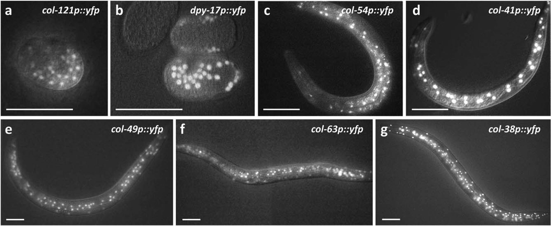
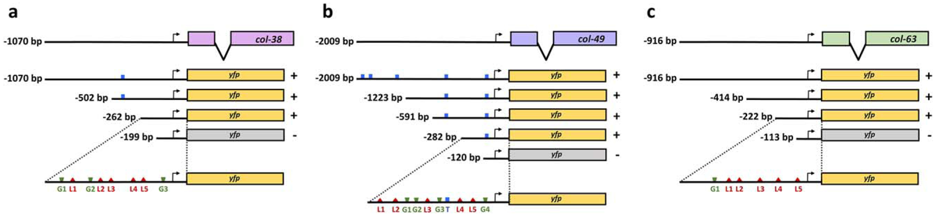
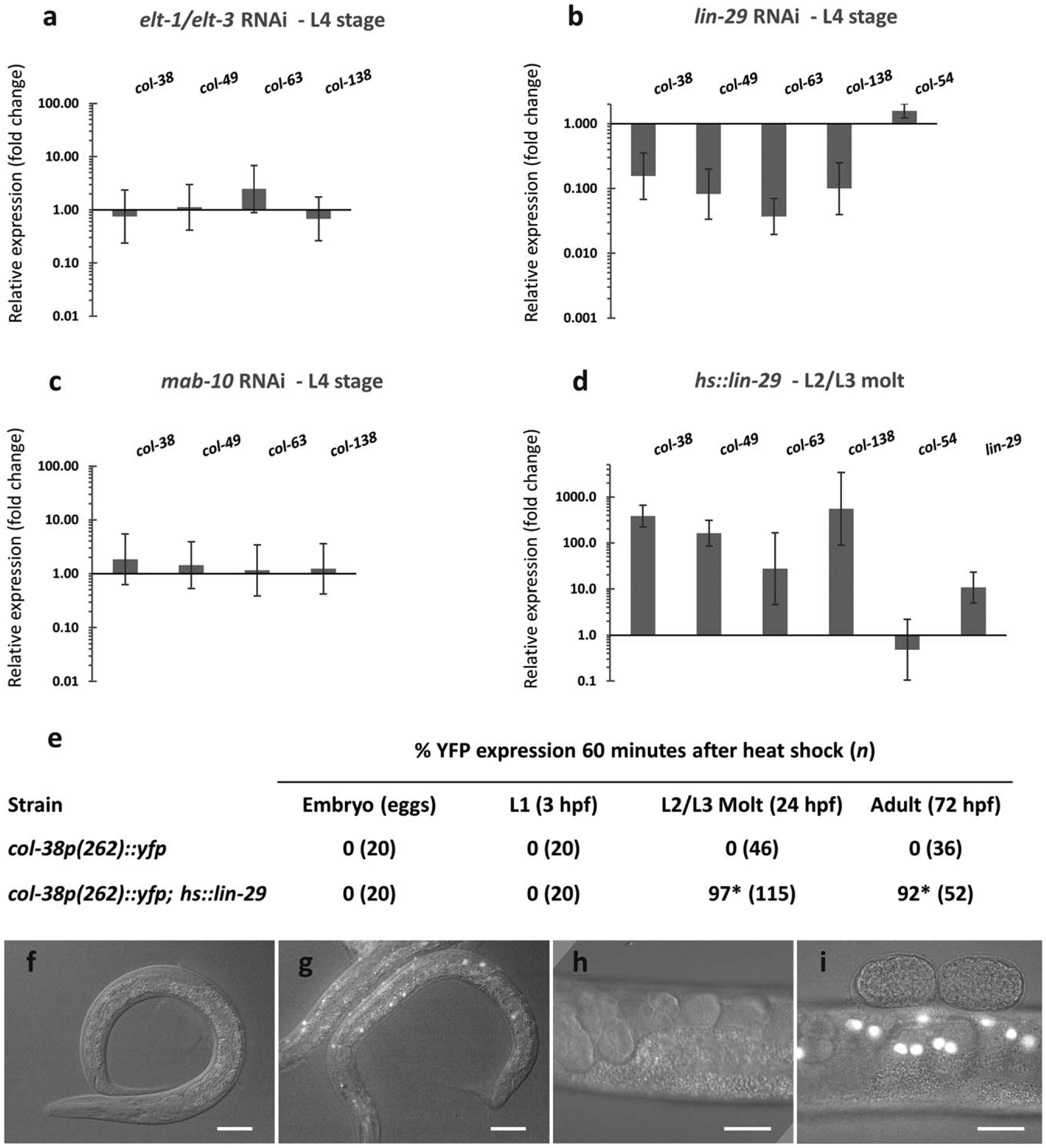
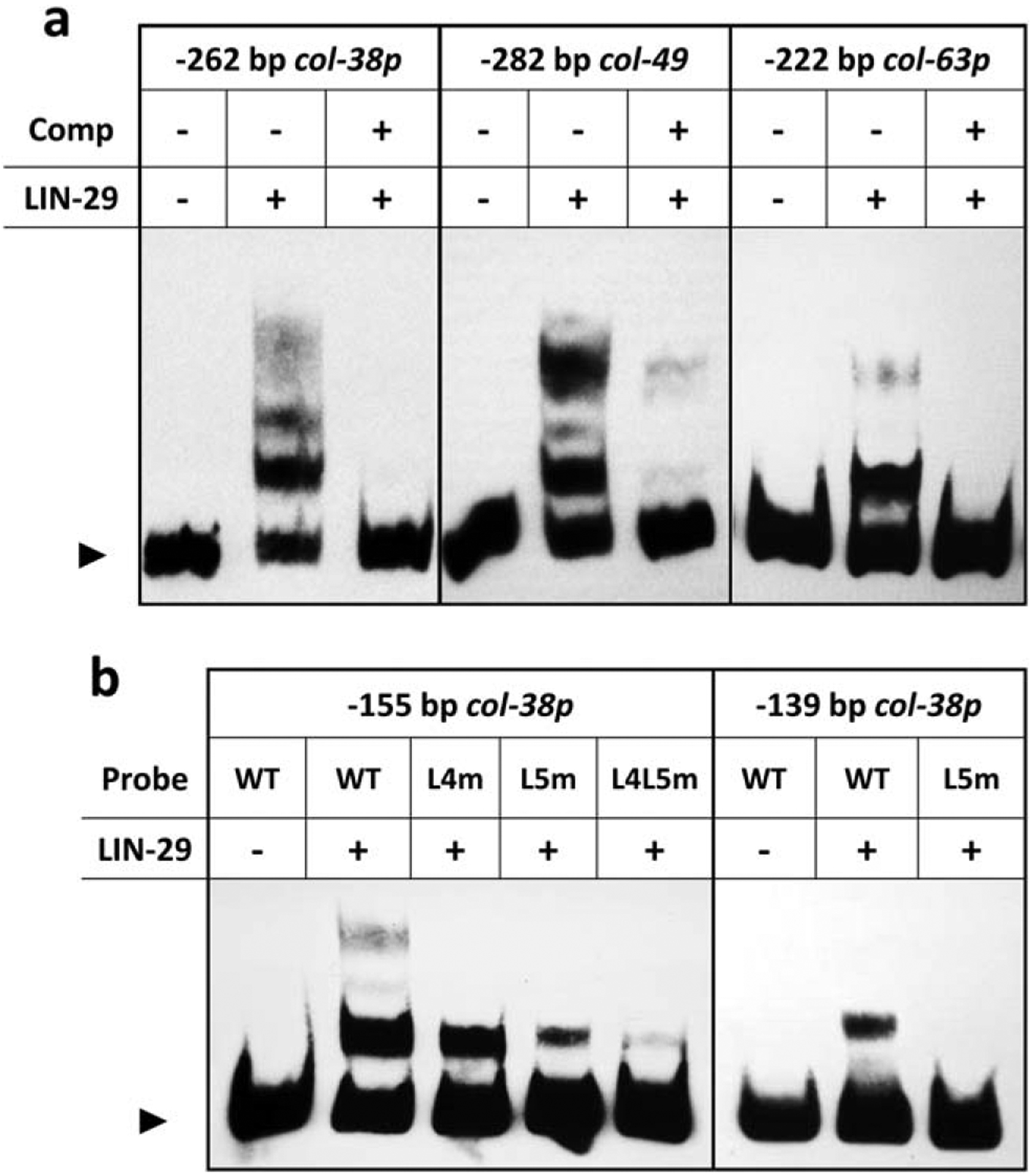
The cuticle, the outer covering of the nematode C. elegans, is synthesized five times during the worm's life by the underlying hypodermis. Cuticle collagens, the major cuticle component, are encoded by a large family of col genes and, interestingly, many of these genes express predominantly at a single developmental stage. This temporal preference motivated us to investigate the mechanisms underlying col gene expression and here we focus on a subset of col genes expressed in the L4 stage. We identified minimal promoter regions of <300 bp for col-38, col-49, and col-63. In these regions, we predicted cis-regulatory sequences and evaluated their function in vivo via mutagenesis of a col-38p::yfp reporter. We used RNAi to study the requirement for candidate transcription regulators ELT-1 and ELT-3, LIN-29, and the LIN-29 co-factor MAB-10, and found LIN-29 to be necessary for the expression of four L4-specific genes (col-38, col-49, col-63, and col-138). Temporal misexpression of LIN-29 was also sufficient to activate these genes at a different developmental stage. The LIN-29 DNA-binding domain bound the col-38, col-49, and col-63 minimal promoters in vitro. For col-38 we showed that the LIN-29 sites necessary for reporter expression in vivo are also bound in vitro: this is the first identification of specific binding sites for LIN-29 necessary for in vivo target gene expression.

Keywords: development; nematode; temporal expression.


Figures

FIGURE 1
FIGURE 1
Stage-specific expression of cuticle collagen genes. Animals carrying the indicated YFP transcriptional reporters were imaged by epifluorescence and Nomarski microscopy at different stages: (a, b) embryo; (c, d) L2 stage; and (e–g) L4 stage. For each reporter, the figure shows the earliest timepoint when YFP was visible during development. YFP expression often perdured past this time. In the case of L2 and L4 stage col reporters, YFP was observed in hypodermal cells of the tail and head, hyp7, and seam cells. For details of col-38p::yfp developmental expression, see Methods. Scale bars indicate 50 μm
FIGURE 2
FIGURE 2
Identification of regulatory regions required for L4 expression in three L4 col YFP reporters. Promoter deletion analyses allowed the identification of minimal promoter regions of 262, 282, and 222 bp in (a) col-38, (b) col-49, and (c) col-63, respectively. For each construct ≥22 animals were assessed in at least two independent lines. In all cases, YFP expression in the L4 stage was either present in ≥80% of the animals (+), or undetectable (−), in which case p<0.001 (Fisher’s exact test) when compared to full length promoter. Locations of predicted binding motifs are shown for TCF/POP-1 (T, blue), GATA factors (G, green), and LIN-29 (L, red)
FIGURE 3
FIGURE 3
LIN-29 is necessary and sufficient for L4 col expression. Endogenous col gene expression in the L4 stage was assessed by RT-qPCR after different RNAi treatments: (a) combined GATA factors elt-1/elt-3 RNAi, (b) lin-29 RNAi and (c) mab-10 RNAi. Quantification was relative to expression in animals treated with empty vector RNAi control. (d) L4 col gene expression was evaluated 1 hr after inducing LIN-29 at the L2/L3 molt in a hs::lin-29 background. Quantification was relative to expression in hs::control animals. col-54 peaks in the L2 stage when lin-29 is not normally expressed, and served as a control. Error bars represent standard errors of the mean. (e) Expression of col-38p(−262)::yfp reporter was assessed after ectopic induction of LIN-29 in the embryo, in the L1, at the L2/L3 molt, and in the adult. (f–i) Epifluorescence and Nomarski microscopy of worms carrying either col-38p(−262)::yfp alone (f,h) or in a hs::lin-29 background (g, i) examined at the L2/L3 molt (f, g) and in the adult (h, i). Scale bars are 25 μm. *p < 0.001 (Fisher’s exact test) when compared to strains carrying col-38p(262)::yfp alone
FIGURE 4
FIGURE 4
LIN-29 binds predicted DNA motifs in L4 col promoters in vitro. (a) Electrophoretic mobility shift assays done with LIN-29 DNA binding domain-GST fusion protein and the minimal promoters of col-38, col-49, and col-63 as probes. In each case, the binding was competed away by a 34 bp oligo consisting of the single col-38 LIN-29 site L5 and its flanking sequence (Comp). (b) Electrophoretic mobility shift assays with LIN-29 DNA binding domain-GST fusion protein and either a −155 bp region of col-38p containing predicted LIN-29 sites L4 and L5 (left) or a −139 bp fragment of col-38p containing only site L5 (right). Binding of LIN-29 to these fragments was reduced or abolished when sites L4 and L5 were mutated individually (L4m, L5m) or together (L4L5m). Arrowhead indicates free probe

References

    1. Baugh LR, Wen JC, Hill AA, Slonim DK, Brown EL, & Hunter CP (2005). Synthetic lethal analysis of Caenorhabditis elegans posterior embryonic patterning genes identifies conserved genetic interactions. Genome Biology, 6, R45. 10.1186/gb-2005-6-5-r45 - DOI - PMC - PubMed
    1. Brenner S (1974). The genetics of Caenorhabditis elegans. Genetics, 77, 71–94. Available at: http://www.ncbi.nlm.nih.gov/pubmed/4366476 - PMC - PubMed
    1. Budovskaya YV, Wu K, Southworth LK, Jiang M, Tedesco P, Johnson TE, & Kim SK (2008). An elt-3/elt-5/elt-6 GATA transcription circuit guides aging in C. elegans. Cell, 134, 291–303. 10.1016/j.cell.2008.05.044 - DOI - PMC - PubMed
    1. Chisholm AD, & Hsiao TI (2012). The C. elegans epidermis as a model skin. I: Development, patterning, and growth. Wiley Interdisciplinary Reviews: Developmental Biology, 1, 861–878. 10.1002/wdev.79 - DOI - PMC - PubMed
    1. Cox GN (1992). Molecular and biochemical aspects of nematode collagens. Journal of Parasitology, 78, 1–15. Retrieved from http://www.jstor.org/stable/3283678 - PubMed

Publication types

MeSH terms

LinkOut - more resources