. 2018 Dec 1;444 Suppl 1(Suppl 1):S274-S286.
doi: 10.1016/j.ydbio.2018.03.022. Epub 2018 Mar 29.

Requirement of zebrafish pcdh10a and pcdh10b in melanocyte precursor migration

Affiliations

Requirement of zebrafish pcdh10a and pcdh10b in melanocyte precursor migration

Jason S Williams et al. Dev Biol. .

Abstract

Melanocytes derive from neural crest cells, which are a highly migratory population of cells that play an important role in pigmentation of the skin and epidermal appendages. In most vertebrates, melanocyte precursor cells migrate solely along the dorsolateral pathway to populate the skin. However, zebrafish melanocyte precursors also migrate along the ventromedial pathway, in route to the yolk, where they interact with other neural crest derivative populations. Here, we demonstrate the requirement for zebrafish paralogs pcdh10a and pcdh10b in zebrafish melanocyte precursor migration. pcdh10a and pcdh10b are expressed in a subset of melanocyte precursor and somatic cells respectively, and knockdown and TALEN mediated gene disruption of pcdh10a results in aberrant migration of melanocyte precursors resulting in fully melanized melanocytes that differentiate precociously in the ventromedial pathway. Live cell imaging analysis demonstrates that loss of pchd10a results in a reduction of directed cell migration of melanocyte precursors, caused by both increased adhesion and a loss of cell-cell contact with other migratory neural crest cells. Also, we determined that the paralog pcdh10b is upregulated and can compensate for the genetic loss of pcdh10a. Disruption of pcdh10b alone by CRISPR mutagenesis results in somite defects, while the loss of both paralogs results in enhanced migratory melanocyte precursor phenotype and embryonic lethality. These results reveal a novel role for pcdh10a and pcdh10b in zebrafish melanocyte precursor migration and suggest that pcdh10 paralogs potentially interact for proper transient migration along the ventromedial pathway.

Keywords: Cell migration; Melanocytes; Neural crest; Protocadherin 10; Zebrafish.


Figures

Figure 1
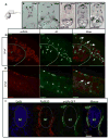
Figure 1. pcdh10a is expressed in a subset of crestin positive migratory neural crest cells (NCCs)
(A–G) Whole-mount in situ hybridization (ISH) of pcdh10a and crestin expression during NCC migration in wildtype zebrafish embryos. (A) pcdh10a is expressed in a similar pattern to the NCC marker crestin (B) Dorsal views at 18 somites. (C,D) Lateral views at 18 somites. (E, F) Lateral views at 24 hpf. (G) Lateral view at 28 hpf. (H, I, K) Confocal micrograph Z-stack projections of double Fluorescent ISH (FISH) at 23hpf and 26 hpf respectively in wildtype zebrafish embryos coexpressing pcdh10a (Red) and crestin (Green). Lateral views with dorsal top, ventral down. White arrows mark examples of colocalization. Scale bars are 50μm. (J) Area of imaging for H, I, K, L. (K) Representative image of a 12μm cross-section of the embryo in (I). White arrows point to areas of colocalization, and white arrowheads point to NCCs in the dorsolateral pathway. (L) 12μm cross-section through the trunk of an embryo stained for Pcdh10 (Red) immunofluorescence, with neural crest cells expressing sox10:EGFP (Green). Scale bar is 20μm, images are a single 1μm Z-plane. NT; Neural Tube, n; Notochord. Anterior is to the left in all whole mount images.
Figure 2
Figure 2. pcdh10a is expressed in migratory NCC-derived melanocyte precursors
(A) Schematic for imaging region for B–I. (B–F) Cross-sectional analysis (12μm) of ISH comparing the melanocyte marker dct with pcdh10a temporally represented across trunk NCC migration time points. (B, C) dct expression in wildtype embryos at 24 hpf and 36 hpf respectively. (D–F) pcdh10a expression in wildtype embryos at 24 hpf, 28 hpf, and 36 hpf respectively. The expression of pcdh10a mirrors that of dct during melanocyte precursor migration suggesting pcdh10a is expressed in melanoblasts. (G, H) Lateral view of whole embryo double FISH comparing expression and colocalization of pcdh10a (red) with dct (green). White arrows are examples of colocalization. Scale bars are 50μm. Single Z-plane images. (I) 12μm cross sections through the embryo trunk stained for Pcdh10 immunofluorescence. Scale bar is 20μm, images are a single 1μm Z-plane, scale bar is 20μm. NT is neural tube, n is notochord. Anterior is to the left in G–H.
Figure 3
Figure 3. Reduction of pcdh10a results in defects in melanoblast migration
(A–D) Lateral views of 72hpf embryos. Line represents approximate position of cross-section. (A) Wildtype embryos with normal larval pigment pattern and all four stripes present, with no internal pigment cells. (B) Schematic showing the difference in morphology and placement of internal pigment cells vs the other correctly patterned stripes (C) Embryos injected with 6ng pcdh10a Morpholino containing several internal pigment cells. (D) pcdh10a −/− embryos also contain internal pigment cells. (A′–D′) cross-sectional views of a representational embryo from A-C. (E) AB embryos injected with 6ng standard control Morpholino show no internal pigment cells. (F) ISH at 25hpf comparing wildtype embryo pcdh10a expression to pcdh10/− mutant pcdh10a expression. (G) Quantification of the number of embryos with at least one internal pigment cell. (H) Quantification of the number of melanocytes at the lateral stripe on both sides of embryo. (I) Table, quantification of the ratio between number of internal melanocytes vs number of lateral stripe melanocytes. DS. Dorsal Stripe, LS. Lateral Stripe, VS. Ventral Strip, YS. Yolk Strip, NT , Neural Tube, n, Notochord Y, Yolk. Arrows show locations of example internal pigment cells. ns, Not Significant
Figure 4
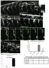
Figure 4. pcdh10a knockdown causes defects in NCC migration
(A–D, F) Lateral views of double transgenic zebrafish Tg(sox10:mRFP,mitfa:GFP), membrane RFP has been changed to gray scale. (A) Control non-injected NCCs 28hpf. (B,C) Lateral view of pcdh10a morphant NCCs at 28hpf show defects in NCC migration, including in mitfa+ melanoblasts. White arrows mark NCCs that have detached from other NCCs in the migrating streams. Red arrows mark NCCs that have aggregated within the neural tube domain. (D) Lateral view at 28hpf of embryos injected with Standard Control MO. Scale bars are 120μm. (E) Time series showing the detachment of a neural crest cells in pcdh10a MO injected embryos, scale bars are 10μm. (F) pcdh10a/−Tg(sox10:mRFP,mitfa:GFP) embryo with subtle NCC aggregations (red arrows). (G) Quantification of the duration of cell detachment events across all samples. (H) Percent of total time NCCs are detached from other NCCs in Control and morphant embryos.(I) Table, comparison of the number of embryos imaged and the number of detachment events that occurred across all samples. * denotes P value of .0062
Figure 5
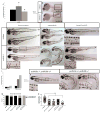
Figure 5. pcdh10b is up-regulated and genetic compensates in pcdh10a mutant embryos
(A) Real-time PCR comparing wildtype embryos to pcdh10a/− mutants, normalized to ef1a, * denotes P-Value <.05. (B–C) ISH probing for pcdh10b expression in (B) wildtype and (C) pcdh10a/− mutants. (D–I) Whole-mount lateral views of 72hpf embryos. Wildtype embryos with no Morpholino injected (D), 2ng pcdh10b Morpholino injected (E), Standard control Morpholino injected (F). (G,H,I) Homozygous pcdh10a/− mutants. With no Morpholino injected (G), 2ng Morpholino injected (H), Standard control Morpholino injected (I). Insets are blown up images to highlight internal melanocytes (red arrows) or lack of internal melanocytes. (J) Mortality rate percent of wildtype embryos and mutant embryo injected with pcdh10b Morpholino. (K, L) Representative images of sibling embryos at 72hpf from the same cross. (K) pcdh10a−/− embryo that is wildtype for pcdh10b. (L) Embryos that are homozygous mutants for both pcdh10a and pcdh10b paralogs. Insets are blown up images to highlight internal melanocytes (red arrows). (M) Quantification of the percent of embryos with internal pigment cells., (N) Quantification of the number of melanocytes in the lateral stripe, **** denotes P-value of 0.0001. (M, N) wildtype n=80, AB 1ng pcdh10a MO n=59, AB 2ng pcdh10a MO n=39, pcdh10a/− 1ng pcdh10b MO n= 113, pcdh10a/, 2ng pcdh10b MO n=22.

Similar articles

Cited by

References

    1. Baker KE, Parker R. Nonsense-mediated mRNA decay: terminating erroneous gene expression. Current Opinion in Cell Biology. 2004;16:293–299. - PubMed
    1. Baxter LL, Moreland RT, Nguyen AD, Wolfsberg TG, Pavan WJ. A curated online resource for SOX10 and pigment cell molecular genetic pathways. Database (Oxford) 2010;2010:baq025. - PMC - PubMed
    1. Bedell VM, Wang Y, Campbell JM, Poshusta TL, Starker CG, Krug RG, 2nd, Tan W, Penheiter SG, Ma AC, Leung AY, Fahrenkrug SC, Carlson DF, Voytas DF, Clark KJ, Essner JJ, Ekker SC. In vivo genome editing using a high-efficiency TALEN system. Nature. 2012;491:114–118. - PMC - PubMed
    1. Belmadani A, Jung H, Ren D, Miller RJ. The chemokine SDF-1/CXCL12 regulates the migration of melanocyte progenitors in mouse hair follicles. Differentiation. 2009;77:395–411. - PMC - PubMed
    1. Bertrand KC, Mack SC, Northcott PA, Garzia L, Dubuc A, Pfister SM, Rutka JT, Weiss WA, Taylor MD. PCDH10 is a candidate tumour suppressor gene in medulloblastoma. Childs Nerv Syst. 2011;27:1243–1249. - PubMed

Publication types

LinkOut - more resources