

Characterizing highly dynamic conformational states: The transcription bubble in RNAP-promoter open complex as an example
- PMID: 29604842
- PMCID: PMC5750056
- DOI: 10.1063/1.5004606
Characterizing highly dynamic conformational states: The transcription bubble in RNAP-promoter open complex as an example
Abstract
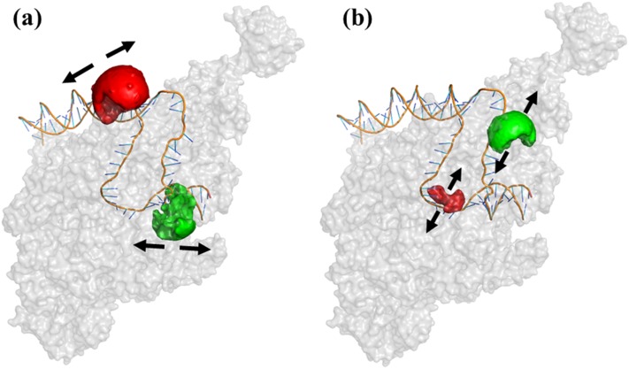
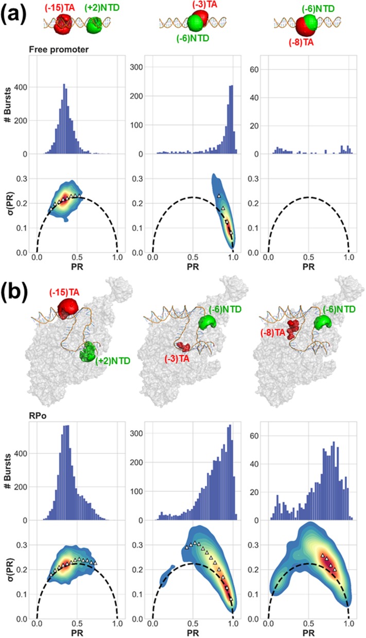
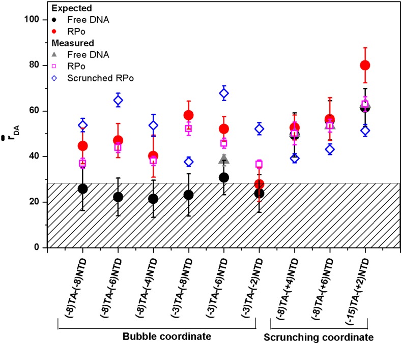
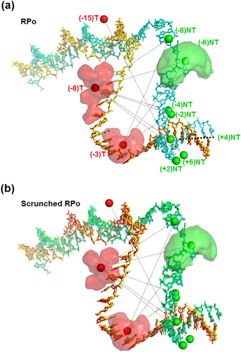
Bio-macromolecules carry out complicated functions through structural changes. To understand their mechanism of action, the structure of each step has to be characterized. While classical structural biology techniques allow the characterization of a few "structural snapshots" along the enzymatic cycle (usually of stable conformations), they do not cover all (and often fast interconverting) structures in the ensemble, where each may play an important functional role. Recently, several groups have demonstrated that structures of different conformations in solution could be solved by measuring multiple distances between different pairs of residues using single-molecule Förster resonance energy transfer (smFRET) and using them as constrains for hybrid/integrative structural modeling. However, this approach is limited in cases where the conformational dynamics is faster than the technique's temporal resolution. In this study, we combine existing tools that elucidate sub-millisecond conformational dynamics together with hybrid/integrative structural modeling to study the conformational states of the transcription bubble in the bacterial RNA polymerase-promoter open complex (RPo). We measured microsecond alternating laser excitation-smFRET of differently labeled lacCONS promoter dsDNA constructs. We used a combination of burst variance analysis, photon-by-photon hidden Markov modeling, and the FRET-restrained positioning and screening approach to identify two conformational states for RPo. The experimentally derived distances of one conformational state match the known crystal structure of bacterial RPo. The experimentally derived distances of the other conformational state have characteristics of a scrunched RPo. These findings support the hypothesis that sub-millisecond dynamics in the transcription bubble are responsible for transcription start site selection.
Figures
Similar articles
-
The transcription bubble of the RNA polymerase-promoter open complex exhibits conformational heterogeneity and millisecond-scale dynamics: implications for transcription start-site selection.J Mol Biol. 2013 Mar 11;425(5):875-85. doi: 10.1016/j.jmb.2012.12.015. Epub 2012 Dec 28. J Mol Biol. 2013. PMID: 23274143 Free PMC article.
-
Stepwise Promoter Melting by Bacterial RNA Polymerase.Mol Cell. 2020 Apr 16;78(2):275-288.e6. doi: 10.1016/j.molcel.2020.02.017. Epub 2020 Mar 10. Mol Cell. 2020. PMID: 32160514 Free PMC article.
-
Structural origins of Escherichia coli RNA polymerase open promoter complex stability.Proc Natl Acad Sci U S A. 2021 Oct 5;118(40):e2112877118. doi: 10.1073/pnas.2112877118. Proc Natl Acad Sci U S A. 2021. PMID: 34599106 Free PMC article.
-
How to switch the motor on: RNA polymerase initiation steps at the single-molecule level.Protein Sci. 2017 Jul;26(7):1303-1313. doi: 10.1002/pro.3183. Epub 2017 May 12. Protein Sci. 2017. PMID: 28470684 Free PMC article. Review.
-
Fluorescence resonance energy transfer analysis of escherichia coli RNA polymerase and polymerase-DNA complexes.Biopolymers. 2001-2002;61(3):201-13. doi: 10.1002/bip.10139. Biopolymers. 2001. PMID: 11987181 Review.
Cited by
-
Ligand Modulates Cross-Coupling between Riboswitch Folding and Transcriptional Pausing.Mol Cell. 2018 Nov 1;72(3):541-552.e6. doi: 10.1016/j.molcel.2018.08.046. Mol Cell. 2018. PMID: 30388413 Free PMC article.
-
Identification and quantification of within-burst dynamics in singly labeled single-molecule fluorescence lifetime experiments.Biophys Rep (N Y). 2022 Sep 14;2(3):100071. doi: 10.1016/j.bpr.2022.100071. Epub 2022 Aug 17. Biophys Rep (N Y). 2022. PMID: 36204594 Free PMC article.
-
Single-photon smFRET. III. Application to pulsed illumination.Biophys Rep (N Y). 2022 Nov 25;2(4):100088. doi: 10.1016/j.bpr.2022.100088. eCollection 2022 Dec 14. Biophys Rep (N Y). 2022. PMID: 36530182 Free PMC article.
-
Multi-parameter photon-by-photon hidden Markov modeling.Nat Commun. 2022 Feb 22;13(1):1000. doi: 10.1038/s41467-022-28632-x. Nat Commun. 2022. PMID: 35194038 Free PMC article.
-
The NEOtrap - en route with a new single-molecule technique.iScience. 2021 Sep 25;24(10):103007. doi: 10.1016/j.isci.2021.103007. eCollection 2021 Oct 22. iScience. 2021. PMID: 34755079 Free PMC article. Review.
References
-
- Brunger A. T., Nat. Struct. Biol. 4 Suppl, 862 (1997). - PubMed
MeSH terms
Substances
Grants and funding
LinkOut - more resources
Full Text Sources
Other Literature Sources