

A fluid membrane enhances the velocity of cargo transport by small teams of kinesin-1
- PMID: 29604873
- PMCID: PMC6910576
- DOI: 10.1063/1.5006806
A fluid membrane enhances the velocity of cargo transport by small teams of kinesin-1
Abstract
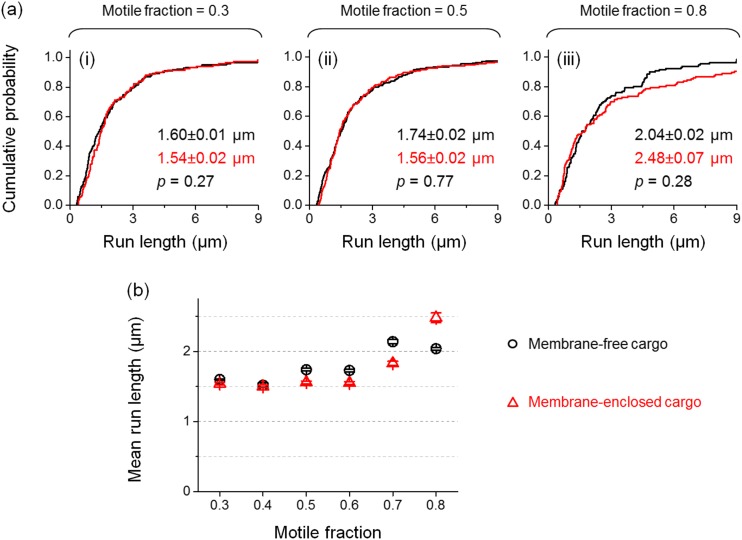
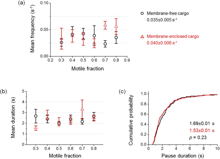
Kinesin-1 (hereafter referred to as kinesin) is a major microtubule-based motor protein for plus-end-directed intracellular transport in live cells. While the single-molecule functions of kinesin are well characterized, the physiologically relevant transport of membranous cargos by small teams of kinesins remains poorly understood. A key experimental challenge remains in the quantitative control of the number of motors driving transport. Here we utilized "motile fraction" to overcome this challenge and experimentally accessed transport by a single kinesin through the physiologically relevant transport by a small team of kinesins. We used a fluid lipid bilayer to model the cellular membrane in vitro and employed optical trapping to quantify the transport of membrane-enclosed cargos versus traditional membrane-free cargos under identical conditions. We found that coupling motors via a fluid membrane significantly enhances the velocity of cargo transport by small teams of kinesins. Importantly, enclosing a cargo in a fluid lipid membrane did not impact single-kinesin transport, indicating that membrane-dependent velocity enhancement for team-based transport arises from altered interactions between kinesins. Our study demonstrates that membrane-based coupling between motors is a key determinant of kinesin-based transport. Enhanced velocity may be critical for fast delivery of cargos in live cells.
Figures


Similar articles
-
Cholesterol in the cargo membrane amplifies tau inhibition of kinesin-1-based transport.Proc Natl Acad Sci U S A. 2023 Jan 17;120(3):e2212507120. doi: 10.1073/pnas.2212507120. Epub 2023 Jan 10. Proc Natl Acad Sci U S A. 2023. PMID: 36626558 Free PMC article.
-
Transport efficiency of membrane-anchored kinesin-1 motors depends on motor density and diffusivity.Proc Natl Acad Sci U S A. 2016 Nov 15;113(46):E7185-E7193. doi: 10.1073/pnas.1611398113. Epub 2016 Nov 1. Proc Natl Acad Sci U S A. 2016. PMID: 27803325 Free PMC article.
-
Two kinesins transport cargo primarily via the action of one motor: implications for intracellular transport.Biophys J. 2010 Nov 3;99(9):2967-77. doi: 10.1016/j.bpj.2010.08.025. Biophys J. 2010. PMID: 21044594 Free PMC article.
-
Kinesin superfamily motor proteins and intracellular transport.Nat Rev Mol Cell Biol. 2009 Oct;10(10):682-96. doi: 10.1038/nrm2774. Nat Rev Mol Cell Biol. 2009. PMID: 19773780 Review.
-
Molecular motor proteins of the kinesin superfamily proteins (KIFs): structure, cargo and disease.J Korean Med Sci. 2004 Feb;19(1):1-7. doi: 10.3346/jkms.2004.19.1.1. J Korean Med Sci. 2004. PMID: 14966333 Free PMC article. Review.
Cited by
-
Methods to Quantify and Relate Axonal Transport Defects to Changes in C. elegans Behavior.Methods Mol Biol. 2022;2431:481-497. doi: 10.1007/978-1-0716-1990-2_26. Methods Mol Biol. 2022. PMID: 35412294
-
Diffusion of kinesin motors on cargo can enhance binding and run lengths during intracellular transport.Mol Biol Cell. 2021 Apr 19;32(9):984-994. doi: 10.1091/mbc.E20-10-0658. Epub 2021 Jan 13. Mol Biol Cell. 2021. PMID: 33439674 Free PMC article.
-
ADP release can explain spatially-dependent kinesin binding times.bioRxiv [Preprint]. 2023 Nov 10:2023.11.08.563482. doi: 10.1101/2023.11.08.563482. bioRxiv. 2023. PMID: 37986962 Free PMC article. Preprint.
-
Cholesterol in the cargo membrane amplifies tau inhibition of kinesin-1-based transport.Proc Natl Acad Sci U S A. 2023 Jan 17;120(3):e2212507120. doi: 10.1073/pnas.2212507120. Epub 2023 Jan 10. Proc Natl Acad Sci U S A. 2023. PMID: 36626558 Free PMC article.
-
Motor Clustering Enhances Kinesin-driven Vesicle Transport.bioRxiv [Preprint]. 2024 Oct 27:2024.10.23.619892. doi: 10.1101/2024.10.23.619892. bioRxiv. 2024. Update in: Biophys J. 2025 Jun 17;124(12):2033-2040. doi: 10.1016/j.bpj.2025.04.033. PMID: 39484389 Free PMC article. Updated. Preprint.
References
MeSH terms
Substances
Grants and funding
LinkOut - more resources
Full Text Sources
Other Literature Sources