

Absence of CFAP69 Causes Male Infertility due to Multiple Morphological Abnormalities of the Flagella in Human and Mouse
- PMID: 29606301
- PMCID: PMC5985338
- DOI: 10.1016/j.ajhg.2018.03.007
Absence of CFAP69 Causes Male Infertility due to Multiple Morphological Abnormalities of the Flagella in Human and Mouse
Abstract
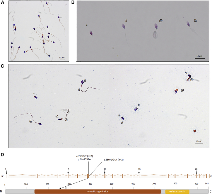
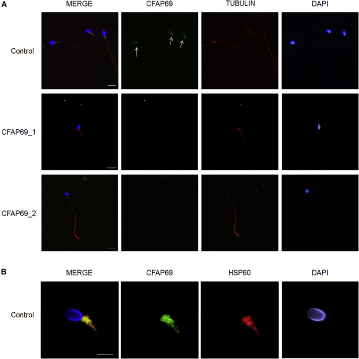
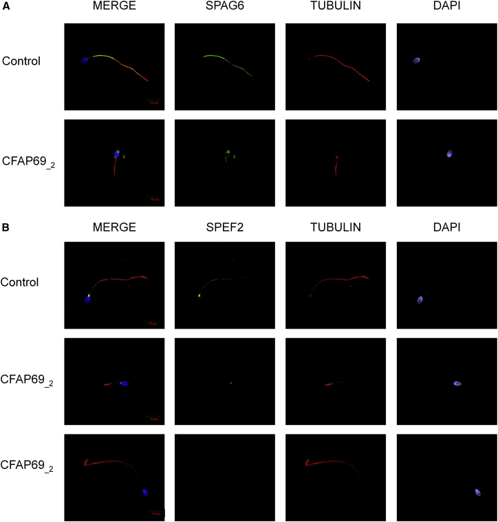
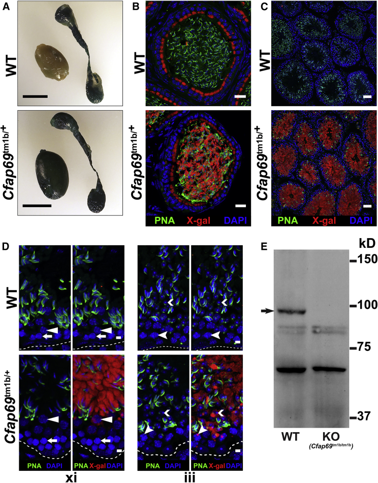
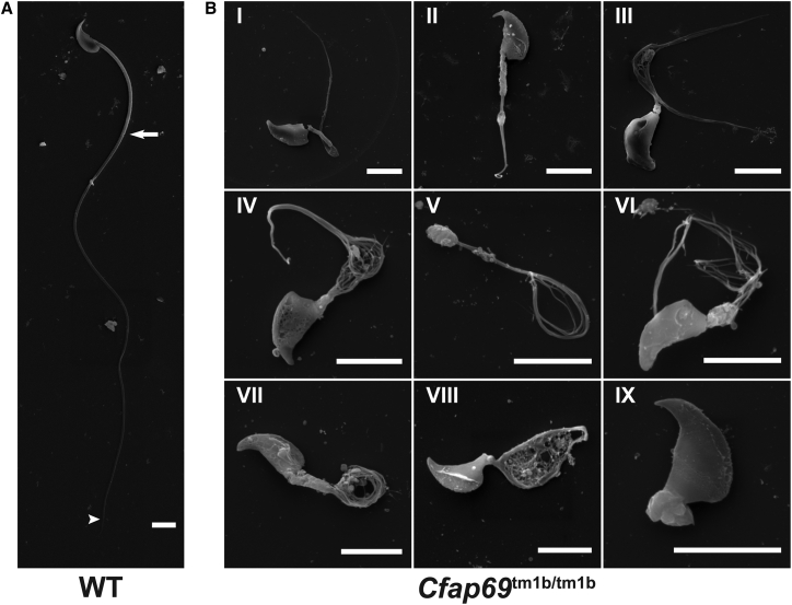
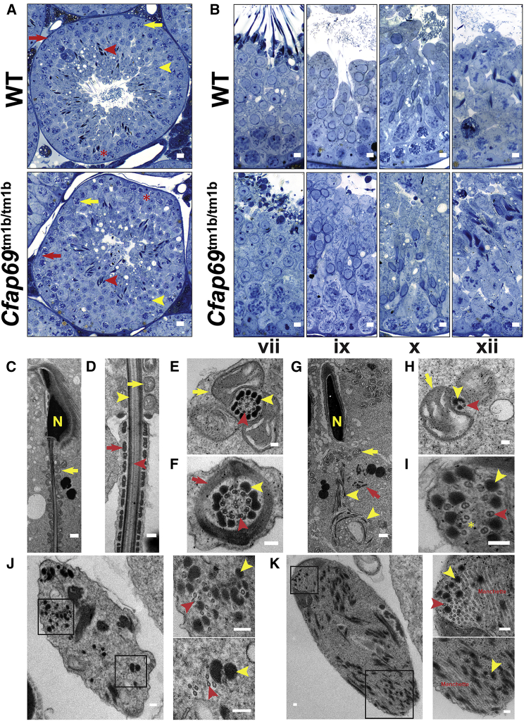
The multiple morphological abnormalities of the flagella (MMAF) phenotype is among the most severe forms of sperm defects responsible for male infertility. The phenotype is characterized by the presence in the ejaculate of immotile spermatozoa with severe flagellar abnormalities including flagella being short, coiled, absent, and of irregular caliber. Recent studies have demonstrated that MMAF is genetically heterogeneous, and genes thus far associated with MMAF account for only one-third of cases. Here we report the identification of homozygous truncating mutations (one stop-gain and one splicing variant) in CFAP69 of two unrelated individuals by whole-exome sequencing of a cohort of 78 infertile men with MMAF. CFAP69 encodes an evolutionarily conserved protein found at high levels in the testis. Immunostaining experiments in sperm from fertile control individuals showed that CFAP69 localized to the midpiece of the flagellum, and the absence of CFAP69 was confirmed in both individuals carrying CFPA69 mutations. Additionally, we found that sperm from a Cfap69 knockout mouse model recapitulated the MMAF phenotype. Ultrastructural analysis of testicular sperm from the knockout mice showed severe disruption of flagellum structure, but histological analysis of testes from these mice revealed the presence of all stages of the seminiferous epithelium, indicating that the overall progression of spermatogenesis is preserved and that the sperm defects likely arise during spermiogenesis. Together, our data indicate that CFAP69 is necessary for flagellum assembly/stability and that in both humans and mice, biallelic truncating mutations in CFAP69 cause autosomal-recessive MMAF and primary male infertility.
Keywords: CFAP69; KO mouse model; MMAF; asthenozoospermia; infertility genetics; male infertility; manchette; sperm flagellum; teratozoospermia; whole-exome sequencing.
Copyright © 2018 American Society of Human Genetics. Published by Elsevier Inc. All rights reserved.
Figures
Similar articles
-
The genetic architecture of morphological abnormalities of the sperm tail.Hum Genet. 2021 Jan;140(1):21-42. doi: 10.1007/s00439-020-02113-x. Epub 2020 Jan 16. Hum Genet. 2021. PMID: 31950240 Review.
-
Novel homozygous CFAP69 mutations in humans and mice cause severe asthenoteratospermia with multiple morphological abnormalities of the sperm flagella.J Med Genet. 2019 Feb;56(2):96-103. doi: 10.1136/jmedgenet-2018-105486. Epub 2018 Nov 10. J Med Genet. 2019. PMID: 30415212
-
Bi-allelic Mutations in ARMC2 Lead to Severe Astheno-Teratozoospermia Due to Sperm Flagellum Malformations in Humans and Mice.Am J Hum Genet. 2019 Feb 7;104(2):331-340. doi: 10.1016/j.ajhg.2018.12.013. Epub 2019 Jan 24. Am J Hum Genet. 2019. PMID: 30686508 Free PMC article.
-
Biallelic Mutations in CFAP43 and CFAP44 Cause Male Infertility with Multiple Morphological Abnormalities of the Sperm Flagella.Am J Hum Genet. 2017 Jun 1;100(6):854-864. doi: 10.1016/j.ajhg.2017.04.012. Epub 2017 May 25. Am J Hum Genet. 2017. PMID: 28552195 Free PMC article.
-
A novel variant in CFAP69 causes asthenoteratozoospermia with treatable ART outcomes and a literature review.J Assist Reprod Genet. 2023 Sep;40(9):2175-2184. doi: 10.1007/s10815-023-02873-1. Epub 2023 Jul 1. J Assist Reprod Genet. 2023. PMID: 37392306 Free PMC article. Review.
Cited by
-
Screening, identification and interaction analysis of key MicroRNAs and genes in Asthenozoospermia.Int J Med Sci. 2021 Feb 6;18(7):1670-1679. doi: 10.7150/ijms.54460. eCollection 2021. Int J Med Sci. 2021. PMID: 33746583 Free PMC article.
-
Ciliary central apparatus structure reveals mechanisms of microtubule patterning.Nat Struct Mol Biol. 2022 May;29(5):483-492. doi: 10.1038/s41594-022-00770-2. Epub 2022 May 16. Nat Struct Mol Biol. 2022. PMID: 35578023 Free PMC article.
-
The genetic architecture of morphological abnormalities of the sperm tail.Hum Genet. 2021 Jan;140(1):21-42. doi: 10.1007/s00439-020-02113-x. Epub 2020 Jan 16. Hum Genet. 2021. PMID: 31950240 Review.
-
Clinical detection, diagnosis and treatment of morphological abnormalities of sperm flagella: A review of literature.Front Genet. 2022 Nov 8;13:1034951. doi: 10.3389/fgene.2022.1034951. eCollection 2022. Front Genet. 2022. PMID: 36425067 Free PMC article. Review.
-
Recent advances and future opportunities to diagnose male infertility.Curr Sex Health Rep. 2019 Dec;11(4):331-341. doi: 10.1007/s11930-019-00225-8. Epub 2019 Oct 26. Curr Sex Health Rep. 2019. PMID: 31853232 Free PMC article.
References
-
- Sharlip I.D., Jarow J.P., Belker A.M., Lipshultz L.I., Sigman M., Thomas A.J., Schlegel P.N., Howards S.S., Nehra A., Damewood M.D. Best practice policies for male infertility. Fertil. Steril. 2002;77:873–882. - PubMed
-
- Ray P.F., Toure A., Metzler-Guillemain C., Mitchell M.J., Arnoult C., Coutton C. Genetic abnormalities leading to qualitative defects of sperm morphology or function. Clin. Genet. 2017;91:217–232. - PubMed
-
- Mitchell M.J., Metzler-Guillemain C., Toure A., Coutton C., Arnoult C., Ray P.F. Single gene defects leading to sperm quantitative anomalies. Clin. Genet. 2017;91:208–216. - PubMed
-
- de Boer P., de Vries M., Ramos L. A mutation study of sperm head shape and motility in the mouse: lessons for the clinic. Andrology. 2015;3:174–202. - PubMed
Publication types
MeSH terms
Substances
Grants and funding
LinkOut - more resources
Full Text Sources
Other Literature Sources
Medical
Molecular Biology Databases
Research Materials
