. 2018 Apr 5;102(4):696-705.
doi: 10.1016/j.ajhg.2018.02.018. Epub 2018 Mar 29.

Bi-allelic Alterations in AEBP1 Lead to Defective Collagen Assembly and Connective Tissue Structure Resulting in a Variant of Ehlers-Danlos Syndrome

Affiliations

Bi-allelic Alterations in AEBP1 Lead to Defective Collagen Assembly and Connective Tissue Structure Resulting in a Variant of Ehlers-Danlos Syndrome

Patrick R Blackburn et al. Am J Hum Genet. .

Abstract

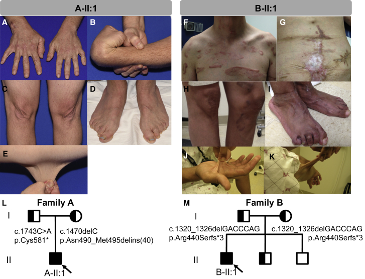
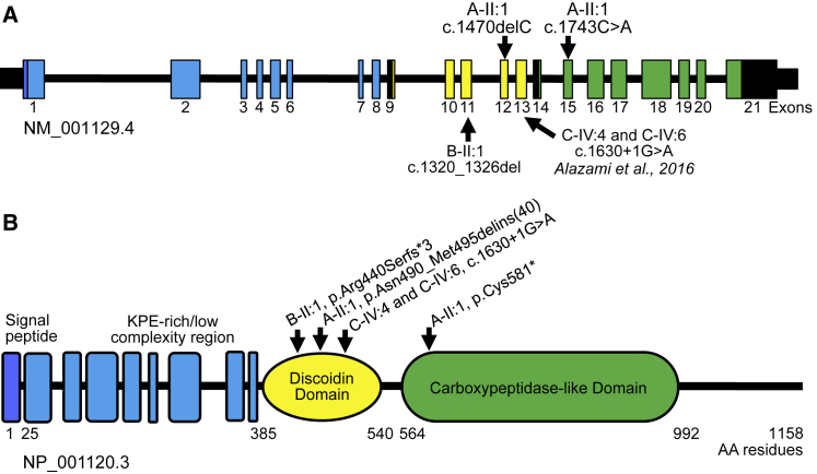
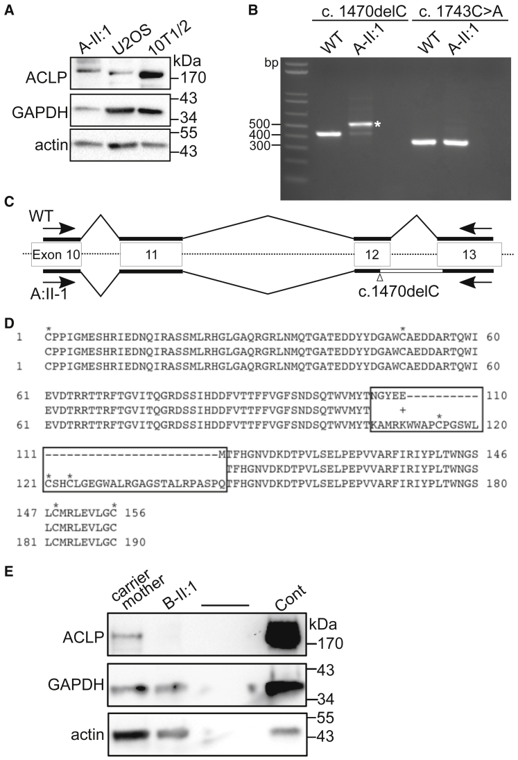
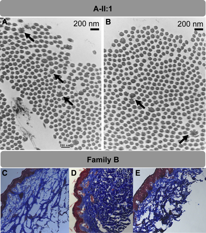
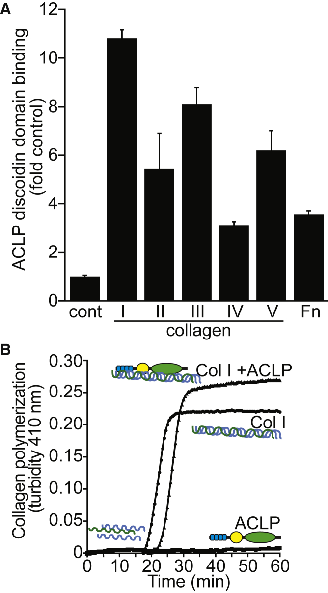
AEBP1 encodes the aortic carboxypeptidase-like protein (ACLP) that associates with collagens in the extracellular matrix (ECM) and has several roles in development, tissue repair, and fibrosis. ACLP is expressed in bone, the vasculature, and dermal tissues and is involved in fibroblast proliferation and mesenchymal stem cell differentiation into collagen-producing cells. Aebp1-/- mice have abnormal, delayed wound repair correlating with defects in fibroblast proliferation. In this study, we describe four individuals from three unrelated families that presented with a unique constellation of clinical findings including joint laxity, redundant and hyperextensible skin, poor wound healing with abnormal scarring, osteoporosis, and other features reminiscent of Ehlers-Danlos syndrome (EDS). Analysis of skin biopsies revealed decreased dermal collagen with abnormal collagen fibrils that were ragged in appearance. Exome sequencing revealed compound heterozygous variants in AEBP1 (c.1470delC [p.Asn490_Met495delins(40)] and c.1743C>A [p.Cys581]) in the first individual, a homozygous variant (c.1320_1326del [p.Arg440Serfs3]) in the second individual, and a homozygous splice site variant (c.1630+1G>A) in two siblings from the third family. We show that ACLP enhances collagen polymerization and binds to several fibrillar collagens via its discoidin domain. These studies support the conclusion that bi-allelic pathogenic variants in AEBP1 are the cause of this autosomal-recessive EDS subtype.

Keywords: ACLP; AEBP1; Aebp1-null mice; Ehlers-Danlos syndrome; aortic carboxypeptidase-like protein; collagen polymerization; connective tissue disorders; discoidin domain; exome sequencing; extracellular matrix.


Figures

Figure 1
Figure 1
Clinical Features of Individuals with AEBP1 Mutations (A–E) Subject A-II:1 at the age of 31 exhibited (A, D) increased wrinkles on his hands and feet with finger contractures, (B) joint hypermobility with shoulder and hip subluxations, (C, E) excess stretchy skin, and (D) bilateral lesser hammertoe and hallux valgus deformities, pes planus, poor wound healing, and abnormal scarring. (F–K) Subject B-II:1 was noted to have dislocated hip that required surgery, easy bruising, and dislocations of his shoulders. He has (F–H) severe abnormal scars that are widened, spread, hyperpigmented, and hypertrophic. At age 27 he presented with a ruptured bowel that was thought to be due to diverticulosis, and underwent repeated attempts to re-anastomose the bowel after which a colostomy bag was required (G). The subject has (I) severe pes planus with hammertoes and hallux valgus deformities as well as (J, K) hyperextensible soft skin. (L) Family pedigree for subject A-II:1 shows that both parents are carriers for nonsense or truncating variants in AEBP1. (M) Family pedigree for subject B-II:1 shows that both parents carry the same small deletion that results in premature truncation in AEBP1. The parents were not consanguineous, but did share some distant ancestry. Half-shaded symbols indicate carrier status and full shading indicates affected individuals in each family. Probands are indicated with arrows.
Figure 2
Figure 2
Schematic Representation of AEBP1, ACLP Protein Structure, and Summary of Known Mutations (A) Exome sequencing revealed compound heterozygous (A-II:1) and homozygous (B-II:1) nonsense and small deletion frameshift variants in AEBP1. The homozygous splice site variant described in two siblings (C-IV:4 and C-IV:6) with similar clinical features by Alazami et al. is also shown. AEBP1 encodes the collagen binding protein. (B) Aortic carboxypeptidase-like protein (ACLP) has several distinct domains that are thought to mediate different protein-protein interactions including a central discoidin domain that helps mediate binding and specificity for fibrillar collagens. ACLP also contains a catalytically inactive metallocarboxypeptidase-like domain. Mutations identified fell within the discoidin domain or immediately downstream and included nonsense, frameshift, and canonical splice site variants that were predicted to result in loss of function.
Figure 3
Figure 3
Analysis of AEBP1 Mutant Fibroblast mRNA and Protein Fibroblasts were isolated from dermal skin biopsies. Cells were routinely cultured in Dulbecco’s Modified Eagle’s Medium (Corning) with 3.7 g/L glucose, 10% fetal bovine serum (Hyclone), and 1% penicillin/streptomycin at 37°C in an 5% CO2 incubator. Cells were passaged before they reached confluence using 0.25% trypsin/EDTA. All cells were genotyped by PCR of genomic DNA and sequencing prior to further analysis. (A) Western blot analysis of protein lysates derived from fibroblasts from subject A-II:1. Anti-ACLP antibodies detected a band >170 kDa. Protein extracts were prepared as previously described, by washing the cells in cold PBS followed by extraction in 25 mM Tris (pH 7.4), 50 mM sodium chloride, 0.5% sodium deoxycholate, 2% NP-40, and 0.2% sodium dodecyl sulfate (SDS) with 1× Complete protease inhibitors cocktail (Roche) and 1× PhosStop phosphatase inhibitors cocktail (Roche). Lysates were incubated on ice for 15 min and cleared by centrifugation at 12,000 × g for 10 min at 4°C. The supernatant was collected and protein concentration was measured using the BCA kit (Thermo Scientific). Protein aliquots were run on 4%–20% Novex SDS-polyacrylamide gels and transferred to nitrocellulose. Blots were probed with antibodies against ACLP and normalized to GAPDH (Sigma G9545, 1:10,000) and pan-actin (Thermo MS-1295B, 1:1,500). (B) Fibroblasts were cultured in 12-well plates and total RNA was isolated and purified using GeneJET RNA Purification Kit (Thermo Scientific). cDNA was obtained using Maxima Reverse Transcriptase (Thermo Scientific). Regions of interest were amplified by PCR using OneTaq DNA polymerase (New England Biolabs) and the following primers: c.1470delC 5′-CCCATTGGGATGGAGTCACA-3′ (forward) 5′-CAGGTGAGTGGGTAGATGCG-3′ (reverse) producing a 422 bp product, and c.1743C>A 5′-GGCTCGTTTCATCCGCATCTA-3′ (forward) 5′-GCACCTCGTTGCCATGGAT-3′ (reverse) producing a 341 bp product. PCR products and controls were run on 2% agarose gels and bands of interest were purified using the QiaQuick Gel Extraction Kit (QIAGEN) followed by sequencing (Eton Bioscience). RNA was extracted from human ACLP-wild-type fibroblasts and from subject A-II:1, and following reverse transcription was amplified with PCR using primers spanning both genomic mutations. A band of approximately 520 bases was amplified (indicated by an asterisk). (C) The DNA fragment (asterisk in B) was purified and subjected to DNA sequencing, which revealed that after the deletion in exon 12, the next intron was retained in the cDNA. (D) The predicted amino acid sequence generated from the results of DNA sequencing and alignment with the wild-type sequence. The boxed region highlights different and additional amino acids, including the addition of three cysteines (indicated by an asterisk). (E) Western blot analysis of protein lysates derived from fibroblasts from subject B-II:1 and his carrier mother with a murine fibroblast control.
Figure 4
Figure 4
Ultrastructural and Histological Analysis of Skin Biopsies For electron microscopy, skin biopsy specimens were fixed in glutaraldehyde, postfixed in osmium tetroxide, stained with uranyl acetate, and embedded in Spurr resin. Thin sections stained with lead citrate were examined on JEOL 1400 transmission electron microscope (JEOL USA). (A and B) Collagen flowers identified by transmission election microscopy (TEM) in the proband in family A. Ultrathin sections of dermal collagen fibrils demonstrates moderate variation in fibril size and scattered composite collagen fibrils (“collagen flowers”); in longitudinal sections, these have a frayed/ragged moth-eaten appearance, and on cross sections, a flower-like appearance. Black arrows point to disordered collagen fibrils. (C–E) Masson’s trichrome staining of skin biopsies from family B show decreased dermal collagen in the proband. (C) B-II:1, the affected proband; (D) carrier brother; (E) carrier mother. Blue, dermis containing collagen bundles; red, epidermis. Original magnification, 100×.
Figure 5
Figure 5
The Discoidin Domain of ACLP Binds to Fibrillar Collagens and Enhances Polymerization of Collagen I In Vitro (A) Binding assays were performed as previously described. Different extracellular matrix proteins were diluted to 10 μg/mL in PBS and each sample was coated to individual wells of a 96-well Corning cell culture plate overnight at room temperature. Proteins used were bovine gelatin (Sigma), rat collagen type I, bovine collagen type II, mouse type III collagen (Fibrogen), mouse collagen type IV, human collagen type V, and human fibronectin (BD Biosciences). Wells were washed with TBST (50 mM Tris [pH 7.4], 150 mM NaCl, 1% Tween-20) and blocked with 1 mg/mL casein (Sigma) in TBST for 1 hr followed by additional washes in TBST. Recombinant anti-Xpress tagged Discoidin-Like-Domain (DLD) protein was purified from BL21 bacteria as previously described. Recombinant DLD binding assays were performed as above with coating of 10 μg/mL of extracellular matrix proteins to a 96-well Corning cell culture plate as described above. Wells were washed in TBST and blocked in 1 mg/mL casein in TBST for 1 hr followed by additional washes in TBST. DLD protein was diluted in TBST to 500 nM and was incubated with each extracellular matrix proteins for 2 hr at room temperature. Wells were washed 3× with TBST and anti-Xpress (Invitrogen) was diluted 1:1,000 in TBST then incubated with each sample for 1 hr. Additional TBST washes were performed and anti-mouse HRP was diluted 1:1,000 in TBST and incubated with each sample for 1 hr followed by washing. Signal was detected using the HRP substrate 3,3′,5,5′-tetramethylbenzidine (TMB) (eBiosciences) and reactions were stopped by the addition of 2 M H2SO4. Plates were read at OD 450 nm and results are presented as either fold control (plastic-only wells). (B) Recombinant ACLP was generated as previously described. In order to explore the role of ACLP in collagen fibril formation, a collagen polymerization assay was used., Collagen I (0.6 mg/mL) was diluted in PBS containing 20 μg/mL rACLP and allowed to polymerize at 37°C for 1 hr in a clear 96-well plate. Readings at 410 nm were taken every 30 s using a BioTek Synergy HT system (Biotek). PBS containing only ACLP was also analyzed as a control.

References

    1. Murphy-Ryan M., Psychogios A., Lindor N.M. Hereditary disorders of connective tissue: a guide to the emerging differential diagnosis. Genet. Med. 2010;12:344–354. - PubMed
    1. Vanakker O., Callewaert B., Malfait F., Coucke P. The genetics of soft connective tissue disorders. Annu. Rev. Genomics Hum. Genet. 2015;16:229–255. - PubMed
    1. Byers P.H., Murray M.L. Heritable collagen disorders: the paradigm of the Ehlers-Danlos syndrome. J. Invest. Dermatol. 2012;132:E6–E11. - PubMed
    1. Zou Y., Donkervoort S., Salo A.M., Foley A.R., Barnes A.M., Hu Y., Makareeva E., Leach M.E., Mohassel P., Dastgir J. P4HA1 mutations cause a unique congenital disorder of connective tissue involving tendon, bone, muscle and the eye. Hum. Mol. Genet. 2017;26:2207–2217. - PMC - PubMed
    1. Hynes R.O., Naba A. Overview of the matrisome--an inventory of extracellular matrix constituents and functions. Cold Spring Harb. Perspect. Biol. 2012;4:a004903. - PMC - PubMed

Publication types

MeSH terms