Review
. 2018 Apr;4(4):292-319.
doi: 10.1016/j.trecan.2018.02.005. Epub 2018 Mar 13.

Reengineering the Physical Microenvironment of Tumors to Improve Drug Delivery and Efficacy: From Mathematical Modeling to Bench to Bedside

Affiliations
Review

Reengineering the Physical Microenvironment of Tumors to Improve Drug Delivery and Efficacy: From Mathematical Modeling to Bench to Bedside

Triantafyllos Stylianopoulos et al. Trends Cancer. 2018 Apr.

Abstract

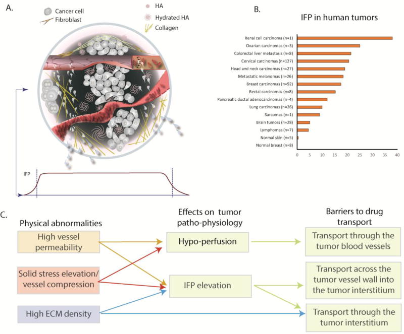
Physical forces have a crucial role in tumor progression and cancer treatment. The application of principles of engineering and physical sciences to oncology has provided powerful insights into the mechanisms by which these forces affect tumor progression and confer resistance to delivery and efficacy of molecular, nano-, cellular, and immuno-medicines. Here, we discuss the mechanics of the solid and fluid components of a tumor, with a focus on how they impede the transport of therapeutic agents and create an abnormal tumor microenvironment (TME) that fuels tumor progression and treatment resistance. We also present strategies to reengineer the TME by normalizing the tumor vasculature and the extracellular matrix (ECM) to improve cancer treatment. Finally, we summarize various mathematical models that have provided insights into the physical barriers to cancer treatment and revealed new strategies to overcome these barriers.


Figures

Figure 1
Figure 1. Abnormalities of the tumor mechanical microenvironment and barriers to drug delivery
(A) Top - A solid tumor consists of cancer and stromal cells, blood and lymphatic vessels and a dense ECM. Blood vessels can be hyper-permeable leading to plasma leakiness and/or collapsed owing to accumulation of solid stress among tumor’s structural components. Collagen deposition and stresses stored in all solid components cause mechanical abnormalities. Bottom: Because of plasma leakage from the hyper-permeable blood vessels, and the loss of lymphatic drainage in the tumor, interstitial fluid pressure (IFP) is elevated throughout the tumor and drops precipitously in the tumor margin (indicated by the dark green color in the schematic). (B) Collective data of IFP measurements in human tumors and normal tissues from published studies.[adapted with permission from [88]] (C) Effects of tumor abnormalities (vessel hyper-permeability, solid stress elevation and high ECM density) on tumor perfusion and IFP and associated barriers to drug delivery. Hypoxia and acidity resulting from poor perfusion can fuel tumor invasion and metastasis and confer resistance to many cancer therapies.
Figure 2
Figure 2. Chronology of measurements of physical parameters in tumors and evolution of mathematical models in our research
(A) Timeline of first measurements of physical and physiological parameters of tumors, their relation to drug delivery in tumors, and strategies to reengineer the tumor microenvironment to improve therapy. (B) Timeline of the evolution of mathematical models throughout our research. Symbols: [a]= [18, 19, 72, 113, 200], [b]=[5, 104, 204], [c]= [, , , –203], [d]= [, –211], [e]=[9, 32, 42], [f]=[12, 55], [g]= [109, 162, 172], [h]= [82, 109, 110, 123, 124], [i]=[162, 213].
Figure 3
Figure 3. Solid mechanics of cancer
(A) Experimental measurements of solid stress from the center towards the tumor periphery. Stress is compressive in at the interior of the tumor, whereas at the periphery becomes tensile. (B) Data showing that solid stress increases as a function of the tumor volume with a lack of increase in matrix stiffness. (C) Tumor mechanical properties between the primary tumor and metastasis are similar in AK4.4 and SL4 tumors but solid stress can differ. (Panels A – C reproduced with permission from [36]). (D) Residual stress is quantifying by the tumor opening, which is the opening that it is formed when a tumor is excised and partially cut along the long axis. Residual stress is evident in all murine and human tumors tested. To account for variations in tumor size, tumor opening is normalized by division with the tumor diameter. (Reproduced with permission from [9]).
Figure 4
Figure 4. Causes, consequences and remedies for solid stress in tumors
(A) Histological data of human tumors show the position of cancer cells (red arrows) and the compression or collapse of blood vessels. Scale bar: 100 µm (reproduced with permission from [12]). (B) Selective depletion of cancer cells, CAFs, collagen or hyaluronan can decrease tumor opening and thus, alleviate solid stress (reproduced with permission from [9]). (C) Tumor perfusion decreases with an increase in tumor volume, presumably owing to accumulation of solid stress (reproduced with permission from [36]).
Figure 5
Figure 5. Fluid mechanics of cancer
(A) Tumor vasculature is irregular and tortuous, lacking a structural hierarchy. The vascular network structure depends on the site of tumor growth (reproduced with permission from [6]). (B) Red blood cell (RBC) velocity in tumors (right) is an order of magnitude lower than that in normal tissues (left) and lacks a correlation with vessel diameter (reproduced with permission from [80]). (C) IFP is elevated and equals microvascular pressure (reproduced with permission from [84]). (D) IFP drops at the tumor periphery resulting in fluid flow from the tumor towards the host tissue. This flow can assist the escape of growth factors, such as VEGF, and metastatic cancer cells fueling tumor progression (reproduced with permission from [20]).
Figure 6
Figure 6. Transvascular and interstitial transport of nanoparticles and macromolecules to tumors
(A) Transvascular flux and penetration of quantum dot particles of hydrodynamic diameters (HD): 12 nm, 60 nm and 125 nm and distinct emission wavelengths, λ. A solution of these nanoparticles was co-injected into mice bearing tumors and their flux through a specific vessel was imaged 120 min after injection (reproduced with permission from [111]). (B) Rods can more effectively extravasate into the tumor compared with spherical particles of the same hydrodynamic diameter (reproduced with permission from [117]). (C) Diffusion coefficient of macromolecules of varying hydrodynamic radius in phosphate buffer saline (PBS) and in U87 glioblastoma implanted in the skin or cranium of immunodeficient mice (reproduced with permission from [108]). Fluorescence image shows the heterogeneous distribution of 90 nm liposomes (bright red color) that are accumulated in perivascular regions (dark color) (reproduced with permission from [116]). (D) Rods can more effectively distribute into the tumor interstitial space compared with spherical nanoparticles of the same hydrodynamic diameter. Intratumor distribution refers to the area of tumor sections occupied by the particles and the distribution of the spherical and rod-like particles in a tumor section (reproduced with permission from [117]).
Figure 7
Figure 7. Vascular normalization strategy
Normalization of tumor vessels using the monoclonal antibody DC101 improves tumor perfusion in a dose-dependent manner. (A) Perfusion images of whole-tumor tissue taken by confocal microscopy. Animals were treated with IgG (control) and 10, 20, or 40 mg/kg of DC101 (green: Sytox staining). Scale bar: 1 mm. (B) Quantification of fractions of Hoechst 33342–positive area in the whole-tumor area presents perfused regions of the tumors shown in panel A for the control (IgG) and the three DC101 doses (D-10, D-20, and D-40). (Reproduced with permission from [133]). (C) Vascular normalization with DC101 or bevacizumab reduces IFP in murine tumor models (reproduced with permission from Ref. [86]). (D) Vascular normalization of murine mammary adenocarcinomas E0771 and 4T1 improves the transvascular transport of nanoparticles in a size-dependent manner (reproduced with permission from [109]). (E) Clinical data of glioblastoma patients who received anti-angiogenic treatment and chemoradiation. Perfusion increased in 20 patients, decreased in 10 patients, and remained stable in 10 patients during combination therapy (left). Kaplan-Meier overall survival data showing that patients whose perfusion was increased exhibited an increased overall survival of ∼ 9 months (Reproduced with permission from [153]).
Figure 8
Figure 8. Stress alleviation strategy
(A) Reengineering the tumor microenvironment with losartan improved the efficacy of the clinically approved cancer nanomedicine Doxil, a liposomal nanoparticle containing doxorubicin (reproduced with permission from [70]). (B) Losartan depleted collagen fibers (blue color) and increased the number of perfused vessels (green color) in preclinical breast tumor models. (C) Combinatorial treatment of losartan with chemotherapy improved overall survival in breast (E0771, 4T1) and pancreatic (AK4.4) tumor models. Scale bar: 1 mm. (reproduced with permission from [71]). (D) Losartan treatment improves outcome in PDAC patients. Previous studies with Folfirinox (FFX) alone or combined with radiation therapy (RT) showed modest improvement in the number of patients that became eligible for surgical resection of the tumor (gray and red bars, studies by Rombouts et al. [221] and Faris et al., [222] respectively). Treatment with losartan combined with chemoradiation therapy (CRT) increased the resection rates dramatically, with 56% of tumors becoming resectable. Remarkably, 52% achieved R0 margins.
Figure I, Text Box 1
Figure I, Text Box 1. Transvascular and interstitial drug transport mechanisms
Transport across the tumor vessel wall and within the tumor interstitial space can be either through diffusion (due to concentration gradients) or convection (due to pressure gradients).

Similar articles

Cited by

References

    1. Jain RK. Normalizing tumor microenvironment to treat cancer: Bench to bedside to biomarkers. J Clin Oncol. 2013;31(17):2205–2218. - PMC - PubMed
    1. Carmeliet P, Jain RK. Angiogenesis in cancer and other diseases. Nature. 2000;407(6801):249–257. - PubMed
    1. Jain RK, et al. Angiogenesis in brain tumours. Nature Reviews Neuroscience. 2007;8(8):610–622. - PubMed
    1. Carmeliet P, Jain RK. Molecular mechanisms and clinical applications of angiogenesis. Nature. 2011;473(7347):298–307. - PMC - PubMed
    1. Gazit Y, et al. Fractal characteristics of tumor vascular architecture during tumor growth and regression. Microcirculation. 1997;4(4):395–402. - PubMed

Publication types

Substances

LinkOut - more resources