. 2018 Apr 11;23(4):470-484.e7.
doi: 10.1016/j.chom.2018.03.004. Epub 2018 Apr 5.

Listeria Adhesion Protein Induces Intestinal Epithelial Barrier Dysfunction for Bacterial Translocation

Affiliations

Listeria Adhesion Protein Induces Intestinal Epithelial Barrier Dysfunction for Bacterial Translocation

Rishi Drolia et al. Cell Host Microbe. .

Abstract

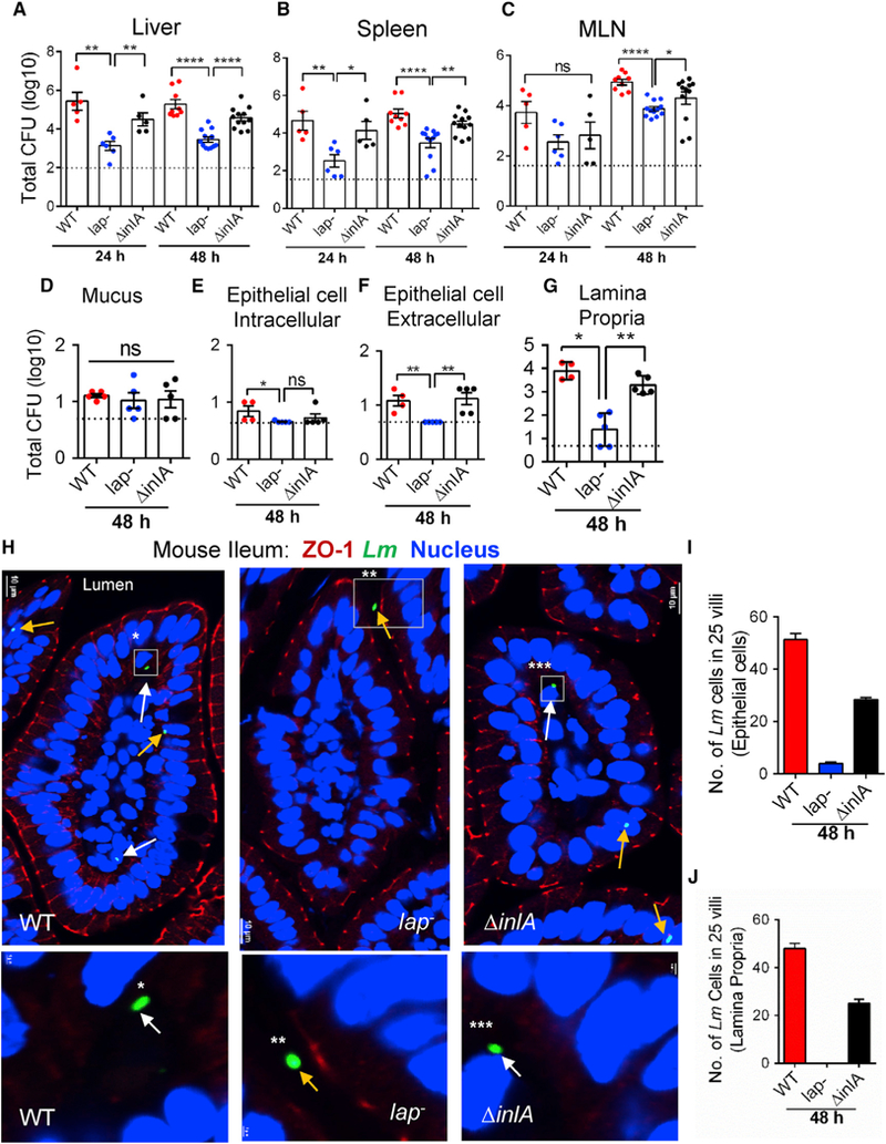
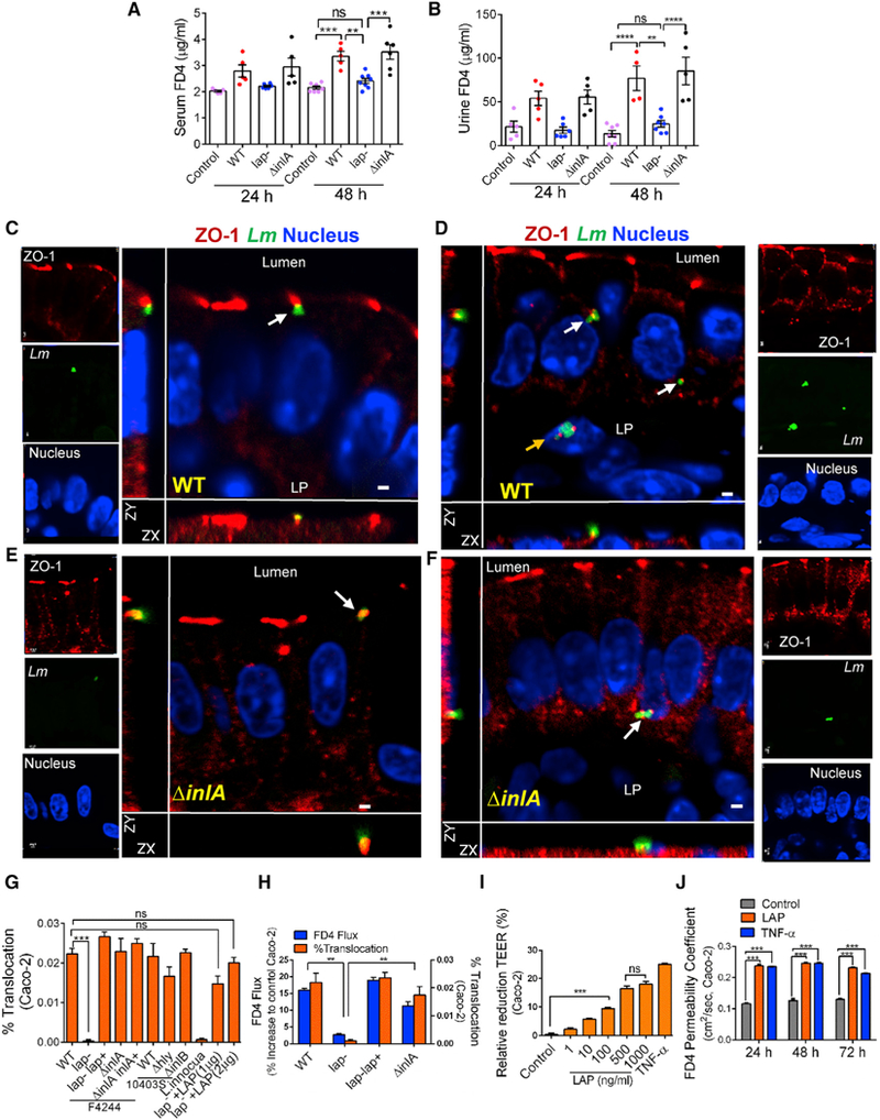
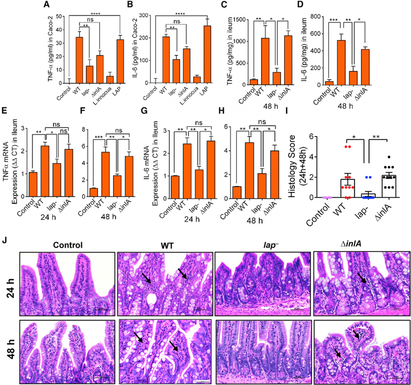
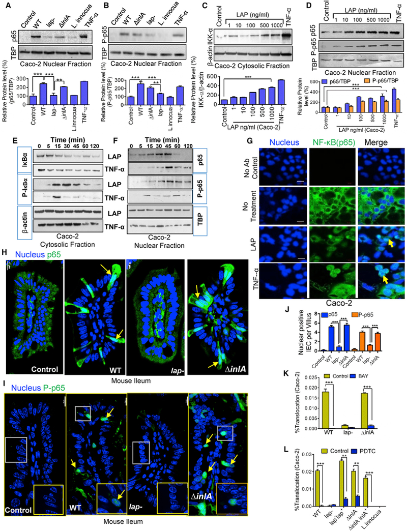
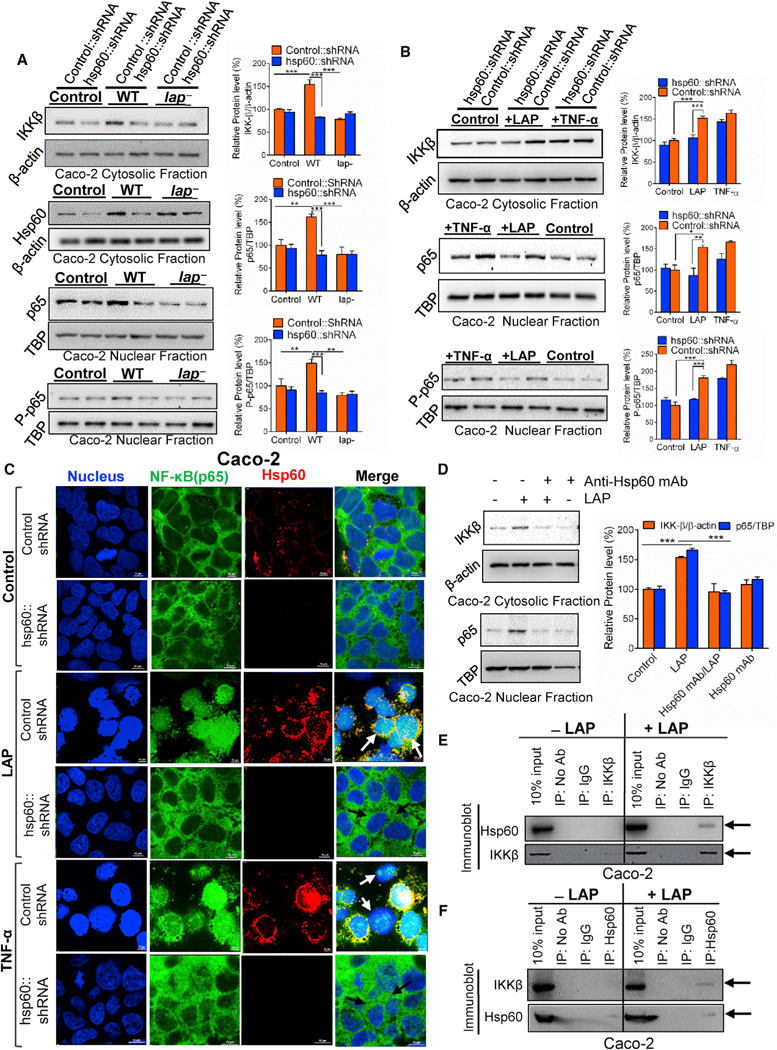
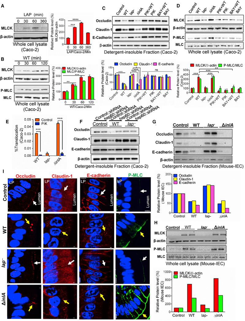
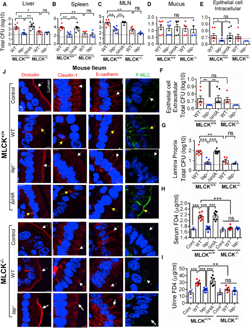
Intestinal epithelial cells are the first line of defense against enteric pathogens, yet bacterial pathogens, such as Listeria monocytogenes, can breach this barrier. We show that Listeria adhesion protein (LAP) induces intestinal epithelial barrier dysfunction to promote bacterial translocation. These disruptions are attributed to the production of pro-inflammatory cytokines TNF-α and IL-6, which is observed in mice challenged with WT and isogenic strains lacking the surface invasion protein Internalin A (ΔinlA), but not a lap- mutant. Additionally, upon engagement of its surface receptor Hsp60, LAP activates canonical NF-κB signaling, facilitating myosin light-chain kinase (MLCK)-mediated opening of the epithelial barrier via cellular redistribution of the epithelial junctional proteins claudin-1, occludin, and E-cadherin. Pharmacological inhibition of MLCK or NF-κB in cells or genetic ablation of MLCK in mice prevents mislocalization of junctional proteins and L. monocytogenes translocation. Thus, L. monocytogenes uses LAP to exploit epithelial defenses and cross the intestinal epithelial barrier.

Keywords: Hsp60; InlA; LAP; Listeria monocytogenes; MLCK; NF-κB; bacterial translocation; intestinal epithelial barrier; mouse; tight junction.


Figures

Figure 1.
Figure 1.. LAP Contributes L. monocytogenes Translocation across the Intestinal Barrier and Systemic Dissemination
(A–G) Listeria counts (Total CFU) in liver (A), spleen (B), MLN (C), ileal mucus (D), epithelial cell intracellular (E), epithelial cell extracellular (F), and lamina propria (G) of A/J mice (n = 4–12) at 24 and 48 hr pi from three independent experiments. Dashed horizontal lines indicate the detection limit. (H) Images of ileal villi (48 hr pi) stained for ZO-1 (red), L. monocytogenes (Lm) (green, arrows), and nucleus (blue). Scale bar, 10 μm. The panels below are enlargements of the boxed areas. Scale bar, 1 μm. White arrows indicate bacteria in the lamina propria and yellow arrows on the epithelial cells or in the lumen. (I and J) Lm counts from epithelial cells (I) and lamina propria (J) of villi images (n = 75) from three mice for each treatment. See also Figure S1. All error bars represent SEM. ****p < 0.0001; **p < 0.01; *p < 0.05; ns, no significance.
Figure 2.
Figure 2.. LAP Contributes to Intestinal Barrier Dysfunction
(A and B) 4-kDa FITC-dextran (FD4) permeability through the intestinal epithelium of uninfected (control) and L. monocytogenes-infected A/J mice in serum (A) and urine (B). (C–F) Lm localization in the mouse ileal epithelial cell junction (48 hr pi). ZO-1 (red), Lm (green, arrows), and nucleus (blue) infected with WT (C and D) or ΔinlA (E and F). Merged images show co-localization of Lm and ZO-1 (arrow), and Lm exiting the epithelial cell (D and F, arrows) and in the lamina propria (LP) (D, yellow arrow). Scale bar, 1 μm. (G and H) L. monocytogenes WT (F4244 and 10403s) and their isogenic mutant strain translocation across Caco-2 cell barrier or lap with exogenously added recombinant LAP (lap +LAP, 1 and 2 μg/mL) and L. innocua (G). (H) FD4 flux (Y1 axis) and bacterial translocation (Y2 axis) after Listeria infection (MOI 50). (I and J) Purified LAP pre-treatment effect on the Caco-2 TEER 48 hr pi (I) and FD4 permeability (J). Human TNF-α (10 ng/mL) was used as a control. See also Figure S2 and Videos S1, S2, S3, S4, and S5. All error bars represent SEM (n = 5–7). ****p < 0.0001; ***p < 0.001; **p < 0.01; ns, no significance.
Figure 3.
Figure 3.. LAP Upregulates TNF-α and IL-6 Expression in Intestinal Cells
(A–D) ELISA analysis of human TNF-α (A) and IL-6 (B) in Caco-2 cell supernatants (n = 4–6), and mouse TNF-α (C) and IL-6 (D) in mouse (n = 3–4) ileal tissues, at 48 hr pi. L. innocua (nonpathogen) and the purified LAP (1 μg/mL) were used as controls (A and B). (E–H) mRNA levels for TNF-α at 24 (E) and 48 hr pi (F) and IL-6 at 24 (G) and 48 hr pi (H) in the ileal mucosa of mice (n = 3–4). Values were normalized to gapdh with the average for untreated samples set at 1. (I and J) Histology score of ileal sections at combined 24 and 48 hr pi (I) and representative hematoxylin and eosin (H&E)-stained images (J) (scale bar, 50 mm) from control uninfected mice (n = 6) or Listeria-infected mice (n = 10–11). Polymorphonuclear and mononuclear cells (arrows) infiltrating the base of the villous lamina propria are evident (J). See also Figure S3 and Table S1. All error bars represent SEM. ****p < 0.0001; ***p < 0.001; **p < 0.01; *p < 0.05; ns, no significance.
Figure 4.
Figure 4.. LAP Contributes to Listeria-Induced NF-κB Activation for Increased Epithelial Permeability
(A and B) Immunoblot and densitometry plots (n = 3) of Listeria-induced p65 (A) and P-p65 (B) levels in the nuclear extracts of uninfected (control) Caco-2 cells or those infected with L. monocytogenes and L. innocua (MOI 50, 30 min). TBP (TATA-binding protein) is a nuclear loading control. (C and D) Immunoblot and densitometry plots (n = 3) showing IKK-α levels in the cytosolic extracts (C) and those of p65 and P-p65 in the nuclear extracts (D) of LAP-treated (30 min) Caco-2 cells. (E and F) Immunoblots showing time-dependent expression of IκBα and P-IκBα in the cytosolic extracts (E) and p65 and P-p65 in the nuclear extracts (F) of Caco-2 cells treated with LAP (1 μg/mL) or TNF-α (10 ng/mL). (G) Images of Caco-2 cells treated with LAP (1 μg/mL) or human TNF-α (10 ng/mL) for 30 min stained for p65 (green) and the nuclei (blue). Arrows show the nuclear localization of p65. Scale bar, 20 μm. (H and I) Images of ileal villi stained forp65 (H, green) and P-p65 (I, green) and the nuclei (blue) from uninfected mice (control) or mice infected with Listeria 48 hr pi (see Figure 1). Arrows indicate the nuclear localization of p65 and P-p65. Scale bar, 10 mm. (J) Enumeration of IECs per villus (n = 10–15 villi from 2–3 mice/treatment) positive for p65 and P-p65 in the nucleus of (H) and (I). (K and L) Listeria (MOI 50) translocation through Caco-2 barrier following pretreatment with BAY (10 μM, 30 min) (K) or PDTC (100 μM, 30 min) (L) (n = 6). See also Figure S4. All error bars represent SEM. ***p < 0.001; **p < 0.01.
Figure 5.
Figure 5.. LAP-Induced NF-kB Activation Is Hsp60 Receptor Dependent
(A and B) Immunoblot and densitometry plots (n = 3) of cytosolic IKK-β and Hsp60 and nuclear p65 and P-p65 In vector control short hairpin RNA (shRNA) (Control::shRNA) or hsp60 knockdown (hsp60::shRNA) Caco-2 cells after infection with Listeria (MOI 50,30 min) or no infection (control) (A). (B) is the same as (A) except the Caco-2 cells were treated with purified LAP (1 μg/mL) or human TNF-α (10 ng/mL) for 30 min (B). (C) Localization of p65 (green), Hsp60 (red), and nucleus (blue) in vector-control shRNA (Control shRNA) or hsp60 knockdown (hsp60::shRNA) Caco-2 cells treated with purified LAP (1 μg/mL) or TNF-α (10 ng/mL) or untreated (control) for 30 min. In LAP and TNF-α-treated control cells, p65 is in the nucleus (white arrows), and it is in the cytoplasm of hsp60 knockdown cells (dark arrows). Scale bar, 10 μm. (D) Immunoblot and densitometry plots (n = 3) of cytosolic IKK-β and nuclear p65 in Caco-2 cells incubated with anti-Hsp60 mAb (1 μg/mL, 1 hr) to block surface Hsp60 prior to LAP (1 μg/mL, 30 min) treatment. (E and F) Immunoblots showing the interaction of Hsp60 with IKK-β in LAP (1 μg/mL, 30 min)-treated Caco-2 cells. IKK-β (E) or Hsp60 (F) was immunoprecipitated from Caco-2 cell lysates and immunoprobed with anti-Hsp60 (E) or anti-IKKβ (F) mAb. Arrows in (E) and (F) indicate co-precipitated IKK-β and Hsp60, respectively, in the LAP-treated cells. The 10% input lane (E and F) represents Caco-2 lysate without immunoprecipitation (IP). The IP: No Ab lane (E and F) represents IP reactions without addition of antibody. Rabbit IgG (IP: IgG lane) (E) or normal mouse IgG (IP: IgG lane) (F) was used asan isotype control IP reaction. See also Figure S5. All error bars represent SEM. ***p < 0.001; **p < 0.01; *p < 0.05.
Figure 6.
Figure 6.. LAP Induces Junctional Protein Dysregulation through MLCK Activation
(A and B) Immunoblot and densitometry plots (n = 3) of MLCK (A and B), P-MLC (B), or MLC (B) in the whole-cell lysate of Caco-2 cells treated with purified LAP (1 μg/mL) (A) or WT (MOI 50) (B). (C and D) Immunoblot and densitometry plots (n = 3) of proteins from the detergent-insoluble fraction (C) and whole-cell lysate of Caco-2 cells infected with Listeria, or pre-treated with the MLCK inhibitor, PIK (150 μM), or the NF-κB inhibitor, BAY (10 μM), prior to infection with WT. (E) Translocation (n = 6) of Listeria (MOI 50, 2 hr) through Caco-2 monolayers pretreated with the MLCK inhibitor, PIK (150 μM). (F) Immunoblots of cell junction proteins in the detergent-insoluble fraction of control or hsp60 knockdown Caco-2 cells infected with Listeria or uninfected (control). (G and H) Immunoblot and densitometry plots of cell junction proteins in the detergent-insoluble fraction (G), and MLCK, P-MLC, and MLC in whole-cell lysates (H) of purified ileal IECs from two mice (A/J) (see Figure 1) at 48 hr pi. (I) Images of the mouse (A/J) ileal villi sections (48 hr pi) showing membrane localization of occludin, claudin-1, E-cadherin (red), and P-MLC (green). White arrows represent normal presentation while yellow arrows represent alterations or mislocalization. Nuclei, blue. Scale bar, 50 μm. LP, lamina propria. See also Figure S6. Caco-2 cell infection (MOI 50, 45 min) (C, D, and F). All error bars represent SEM. ***p < 0.001; **p < 0.01, *p < 0.05; ns, no significance.
Figure 7.
Figure 7.. L. monocytogenes Translocation and Epithelial Permeability Did Not Increase in MLCK Knockout Mice
(A–G) Listeria counts (Total CFU) in liver (A), spleen (B), MLN (C), ileal mucus (D), epithelial cell intracellular (E), epithelial cell extracellular (F), and lamina propria (G) of WT C57BL/6 (MLCK+/+) or MLCK knockout (MLCK−/−) mice (n = 4–7 male and female) at 48 hr pi. Dashed lines indicate the detection limit. (H and I) Analysis of FD4 permeability through the intestinal epithelium of uninfected (Cont) and WT, lap “-infected, and ΔinlA-infected MLCK+/+ or MLCK−/− mice (n = 5–7) in serum (H) and urine (I) at 48 hr pi. (J) Images of the mouse (C57BL/6, MLCK+/+, and MLCK−/−) ileal villi sections (48 hr pi) showing membrane localization of occludin, claudin-1, E-cadherin (red), and P-MLC (green). White arrows represent normal presentation, while yellow arrows represent alterations or mislocalization. Nuclei, blue. Scale bar, 50 μm. LP, lamina propria. All error bars represent SEM. ***p < 0.001; **p < 0.01; *p < 0.05; ns, no significance.

Similar articles

Cited by

References

    1. Abreu MT, Thomas LS, Arnold ET, Lukasek K, Michelsen KS, and Arditi M (2003). TLR signaling at the intestinal epithelial interface. J. Endotoxin Res. 9, 322–330. - PubMed
    1. Baldwin DN, Vanchinathan V, Brown PO, and Theriot JA (2003). Agene-expression program reflecting the innate immune response of cultured intestinal epithelial cells to infection by Listeria monocytogenes. Genome Biol. 4, R2. - PMC - PubMed
    1. Berger E, Rath E, Yuan D, Waldschmitt N, Khaloian S, Allgauer M, Staszewski O, Lobner EM, Schottl T, Giesbertz P, et al. (2016). Mitochondrial function controls intestinal epithelial stemness and proliferation. Nat. Commun. 7, 13171. - PMC - PubMed
    1. Bierne H, Mazmanian SK, Trost M, Pucciarelli MG, Liu G, Dehoux P, Jaansch L, Garcia-del Portillo F, Schneewind O, and Cossart P; European Listeria Genome Consortium (2002). Inactivation of the srtA gene in Listeria monocytogenes inhibits anchoring of surface proteins and affects virulence. Mol. Microbiol. 43, 869–881. - PubMed
    1. Bonazzi M, Veiga E, Pizarro-Cerda J, and Cossart P (2008). Successive post-translational modifications of E-cadherin are required for InlA-mediated internalization of Listeria monocytogenes. Cell. Microbiol. 70, 2208–2222. - PubMed

Publication types