Case Reports
. 2018 Apr;59(4):379-384.

Retrobulbar malignant peripheral nerve sheath tumor in a golden retriever dog: A challenging diagnosis

Affiliations
Case Reports

Retrobulbar malignant peripheral nerve sheath tumor in a golden retriever dog: A challenging diagnosis

Cécile Briffod et al. Can Vet J. 2018 Apr.

Abstract

A 9-year-old golden retriever dog was diagnosed with a left retrobulbar mass. Fine-needle aspirations and incisional biopsies resulted in discordant diagnoses: myxosarcoma/myxoma or rhadomyosarcoma, respectively. Immunohistochemistry following exenteration allowed definitive diagnosis of malignant peripheral nerve sheath tumor with fibromyxomatous differentiation. Fifteen weeks after surgery, an aggressive recurrence resulted in euthanasia.

Tumeur rétrobulbaire maligne des gaines nerveuses périphériques chez un Golden Retriever : un défi diagnostique. Une masse rétrobulbaire gauche a été diagnostiquée chez une Golden Retriever de 9 ans. Des aspirations à l’aiguille fine et des biopsies incisionnelles ont établi des diagnostics discordants : un myxosarcome/myxome ou un rhabdomyosarcome, respectivement. Suite à l’exentération, l’immunohistochimie a permis un diagnostic définitif de tumeur maligne des gaines nerveuses périphériques avec différenciation fibro-myxomateuse. Quinze semaines après la chirurgie, une récidive agressive a conduit à l’euthanasie de la chienne.(Traduit par les auteurs).


Figures
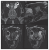
Figure 1
Figure 1
Dorsal (a) and sagittal (b) fast spin echo T2W (TR = 5650 ms and TE = 85 ms) MR images of the head. The left retrobulbar mass is well-defined and highly hyperintense to brain parenchyma. The mass infiltrates the left medial retrobulbar region and is associated with a mass effect creating a compression and a rostrolateral displacement of the globe. Transverse T1W-FLAIR (TR = 2403 ms and TE = 26 ms) (c) pre- and (d) post-contrast MR images. The mass is hypointense and heterogeneous on pre-contrast T1W images (c) but shows strong heterogenous contrast enhancement (d). Note the compression of the left globe on both images. The zygomatic gland appeared normal and unaffected by the process (c — white circle).
Figure 2
Figure 2
a — Light microscopic image from echo-guided biopsy showing a mass with low cellularity with spindle cells in an abundant fibrovascular or myxoid stroma. Note the spindle cell with ill-defined margins, eosinophilic cytoplasm, and elongated nucleus consistent with muscle cells. b — Alcian blue stain showing abundant blue staining of mucin and pink to red staining of cells nuclei consistent with the myxoid feature of the mass.
Figure 3
Figure 3
a — Light microscopic image demonstrating spindle neoplastic cells, arranged in a vaguely storiform pattern, separated by a loose fibromyxomatous matrix. Note the admixed hemosiderophages (arrows) that were also observed during cytologic examination. Inset. An infiltrative retrobulbar neoplasm that compresses the posterior segment (asterisk). Hematoxylin and eosin (H&E). b and c — Immunohistochemical characterization of neoplastic cells. Neoplastic cells show strong nuclear and cytoplasmic immunoreactivity for S100 protein (b) as well as cytoplasmic immunostaining for GFAP (c).
Figure 4
Figure 4
Transverse CT images (a) pre- (bone reconstruction algorithm) and (b) (detail reconstruction algorithm) post-contrast at the level of the caudal retro-orbital space. Note the bone lysis of the medial wall of the left orbit (a). This mass is characterised by multiple fluid pockets with ring enhancement. Note the extension of the mass into the cranial vault and the mass effect on olfactory bulb (b). Dorsal CT image (detail reconstruction algorithm) (c) post-contrast demonstrating invasion of the left nasal cavity.

Similar articles

Cited by

References

    1. Attali-Soussay K, Jegou JP, Clerc B. Retrobulbar tumors in dogs and cats: 25 cases. Vet Ophthalmol. 2001;4:19–27. - PubMed
    1. Hendrix DV, Gelatt KN. Diagnosis, treatment and outcome of orbital neoplasia in dogs: A retrospective study of 44 cases. J Small Anim Pract. 2000;41:105–108. - PubMed
    1. Hagen FV, Romkes G, Kershaw O, Eule JC. Malignant peripheral nerve sheath tumor of the third eyelid in a 3-year-old Rhodesian Ridgeback. Clin Case Reports. 2015;3:50–56. - PMC - PubMed
    1. Sfacteria A, Perillo L, Macrì F, Lanteri G, Rifici C, Mazzullo G. Peripheral nerve sheath tumor invading the nasal cavities of a 6-year-old female Pointer dog. Vet Q. 2015;35:170–173. - PubMed
    1. Curto E, Clode AB, Durrant J, Montgomery KW, Gilger BC. Retrobulbar pigmented peripheral nerve sheath tumor in a dog. Vet Ophthalmol. 2016;19:518–524. - PubMed

Publication types

LinkOut - more resources