Review
. 2018 Mar 22:13:437-449.
doi: 10.2147/CIA.S156615. eCollection 2018.

The reliability of lung ultrasound in assessment of idiopathic pulmonary fibrosis

Affiliations
Review

The reliability of lung ultrasound in assessment of idiopathic pulmonary fibrosis

Diana Manolescu et al. Clin Interv Aging. .

Abstract

Idiopathic pulmonary fibrosis (IPF) is the severest form of idiopathic interstitial pneumonia, with a median survival time estimated at 2-5 years from the time of diagnosis. It occurs mainly in elderly adults, suggesting a strong link between the fibrosis process and aging. Although chest high-resolution computed tomography (HRCT) is currently the method of choice in IPF assessment, diagnostic imaging with typical usual interstitial pneumonia (UIP) provides definitive results in only 55%, requiring an invasive surgical procedure such as lung biopsy or cryobiopsy for the final diagnostic analysis. Lung ultrasound (LUS) as a noninvasive, non-radiating examination is very sensitive to detect subtle changes in the subpleural space. The evidence of diffuse, multiple B-lines defined as vertical, hyperechoic artifacts is the hallmark of interstitial syndrome. A thick, irregular, fragmented pleura line is associated with subpleural fibrotic scars. The total numbers of B-lines are correlated with the extension of pulmonary fibrosis on HRCT, being an LUS marker of severity. The average distance between two adjacent B-lines is an indicator of a particular pattern on HRCT. It is used to appreciate a pure reticular fibrotic pattern as in IPF compared with a predominant ground glass pattern seen in fibrotic nonspecific interstitial pattern. The distribution of the LUS artifacts has a diagnostic value. An upper predominance of multiple B-lines associated with the thickening of pleura line is an LUS feature of an inconsistent UIP pattern, excluding the IPF diagnosis. LUS is a repeatable, totally radiation-free procedure, well tolerated by patients, very sensitive in detecting early changes of fibrotic lung, and therefore a useful imaging technique in monitoring disease progression in the natural course or after initiation of treatment.

Keywords: B-lines artifacts; chest high-resolution computed tomography; chest ultrasound; interstitial lung diseases; interstitial syndrome.


Conflict of interest statement

Disclosure The authors report no conflicts of interest in this work.

Figures

Figure 1
Figure 1
The physical and anatomic basis of echo lung comets. Notes: (A) Normally aerated lung with A-lines artifact as reflection of pleura line. (B) Reflections of the ultrasound beam by the thickened interlobular septa proved comet-tail artifact (B-lines) in patients with IPF. Modified from Am J Cardiol, 93(10), Jambrik Z, Monti S, Coppola V, et al, Usefulness of ultrasound lung comets as a nonradiologic sign of extravascular lung water, 1265–1270, Copyrgiht (2004), with permission from Elsevier. Abbreviation: IPF, idiopathic pulmonary fibrosis.
Figure 2
Figure 2
Aspect of A-lines (arrows) in (A) normal lung, (B) emphysematous lung, and (C) IPF with honeycombing. Abbreviation: IPF, idiopathic pulmonary fibrosis.
Figure 3
Figure 3
HRCT-LUS correlation in normal lung, pulmonary congestion, rheumatoid lung disease, idiopathic pulmonary fibrosis. Notes: (A) Normal lung transparency; (B) A-lines (arrow head) reverberation artifacts from normal interlobular septa; (C) heart failure with extensive ground glass opacities; (D) “white lung” due to alveolar syndrome with multiple long, vertical hyperechoic B-line artifacts (thin arrow) arising from a smooth pleura line (yellow arrow); (E) NSIP fibrotic lung with extensive ground glass opacities and traction bronchiectasis due to pulmonary volume loss; (F) >10 B-lines in one IC with thickened, fragmented pleura line (red arrow) in a fibrotic alveolar syndrome (thin arrow shows B-line artifact); (G) UIP pattern with subpleural, basal reticular abnormalities, traction bronchiectasis, and honeycombing; (H) B-lines with irregular thick pleura line (thin arrows show B-line artifacts). Abbreviations: HRCT, high-resolution computed tomography; LUS, lung ultrasound; NSIP, nonspecific interstitial pattern; IC, intercostal space; UIP, usual interstitial pneumonia; GGO, ground glass opacity.
Figure 4
Figure 4
Pleura line appearance. Notes: (A) Thickened (>3 mm) irregular pleura line due to subpleural fibrotic abnormalities; (B) normal thickness (2 mm).
Figure 5
Figure 5
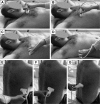
Patient position during LUS, with the probe placed along the (A) paravertebral, (B) midclavicular, (C) anterior axillary, (D) medial axillary, (E) posterior axillary, (F) subscapular, and (G) paravertebral lines. Abbreviation: LUS, lung ultrasound.
Figure 6
Figure 6
LIS assessment in a reduced LUS protocol (14 LIS). 1: Second IC along parasternal line; 2: fourth IC along midclavicular line; 3: fourth IC along anterior axillary line; 4: fourth IC along medial axillary line; 5: eighth IC along posterior axillary line; 6: eighth IC along subscapular line; 7: eighth IC along paravertebral line. Red asterisks represent places of examination. Abbreviations: LIS, lung intercostal spaces; LUS, lung ultrasound; IC, intercostal space.
Figure 7
Figure 7
Correlation of HRCT and LUS of a UIP pattern in IPF. Notes: (A, B) Axial HRCT imaging in the lung window at the level of middle and lower lobes with subpleural reticulation with a basal predominance; honeycombing (black arrows) and traction bronchiectasis (red arrow) associated in >50%. (C, D) LUS at the level of both sixth PV lines: >3 B-lines are seen per IS (white thin arrows) with a median distance between one another of 5.6 mm; irregular, thickened, blurred pleural line confirmed the fibrotic interstitial syndrome. The yellow arrow shows the pleural line. Abbreviations: HRCT, high-resolution computed tomography; LUS, lung ultrasound; NSIP, nonspecific interstitial pattern; IPF, idiopathic pulmonary fibrosis; UIP, usual interstitial pneumonia; PV, paravertebral; IS, intercostal space.
Figure 8
Figure 8
Correlation of HRCT and LUS of an NSIP pattern in a patient with rheumatoid lung disease. Notes: (A) Axial HRCT imaging in the lung window at the level of middle and lower lobes with extensive GGO (black arrow) and fine reticulation; traction bronchiectasis as a mark of lung volume loss. (B, C) LUS at the level of both eighth SS and PV line: “white lung” (thick white arrow) due to very close multiple B-lines (thin white arrows); irregular, thickened, blurred pleural line (yellow arrow) confirmed the fibrotic interstitial syndrome. Abbreviations: HRCT, high-resolution computed tomography; LUS, lung ultrasound; NSIP, nonspecific interstitial pattern; PV, paravertebral; SS, subscapular; GGO, ground glass opacity.
Figure 9
Figure 9
Correlation of HRCT and LUS of an inconsistent UIP pattern in a patient with chronic hypersensitivity pneumonitis. Notes: (AC) Axial HRCT imaging in the lung window at the level of upper, middle, and lower lobes with subpleural reticulation; traction bronchiectasis and mild honeycombing (black arrow). Patchy areas of mosaic attenuation due to small bronchial obstruction (red arrow). (A1, A2) LUS: multiple B-lines (thin arrow), with a thick 3.7 mm pleura line (yellow arrow), are observed in the evaluation of the second IS along PV line on (A1) the right and (A2) the left side; (B1, B2) LUS: B-lines at the fourth IS along MA line on both sides; (C1) LUS: preserved right lung base with thin pleura line and normal A-lines at eighth IS along PV line; (C2) LUS: thin pleura line with a few B-lines and normal A-lines corresponding to mild abnormalities at the eighth IS along SS line. Abbreviations: HRCT, high-resolution computed tomography; LUS, lung ultrasound; UIP, usual interstitial pneumonia; IS, intercostal space; MA, medial axillary; PV, paravertebral; SS, subscapular.
Figure 10
Figure 10
LUS assessment of the thickness of pleura line at the upper (A) versus basal (B) chest regions. Abbreviation: LUS, lung ultrasound.
Figure 11
Figure 11
Correlation of HRCT and LUS in a patient with possible UIP pattern and biopsy-proven idiopathic pulmonary fibrosis. Notes: (AC) Axial HRCT imaging in the lung window at the level of upper, middle, and lower lobes with subpleural reticulation and fine traction bronchiectasis with a basal lung predominance (black arrow). No feature of honeycombing. (A1, A2) LUS at the second IS along MC line: normal aerated lung with A-lines (thin white arrow) and smooth pleura line (curve yellow arrow); (B1, B2) LUS at the level of fourth along MA line: a few B-lines (white arrow head) in the lateral lung regions with an irregular pleura line (yellow arrow); (C1, C2) LUS at the level of eighth along PV line: increased B-line (arrowheads) number with thick, irregular pleura line. Abbreviations: HRCT, high-resolution computed tomography; LUS, lung ultrasound; UIP, usual interstitial pneumonia; IS, intercostal space; MA, medial axillary; PV, paravertebral; MC, midclavicular.
Figure 12
Figure 12
Correlation of HRCT and LUS in a patient with IPF. Notes: (AC) Axial HRCT imaging in the lung window at the level of upper, middle, and lower lobes with moderate to severe UIP pattern defined by extensive subpleural areas of reticulation with honeycombing and traction bronchiectasis. LUS images: bilateral multiple B-lines (arrow head) with thick, irregular, blurred pleura line (yellow arrow) in all zones: (A1, A2) at the level of second IS along MC line; (B1, B2) at the level of sixth IS along MA line; (C1, C2) at the level of eighth PV line. Abbreviations: HRCT, high-resolution computed tomography; LUS, lung ultrasound; UIP, usual interstitial pneumonia; IPF, idiopathic pulmonary fibrosis; IS, intercostal space; MA, medial axillary; PV, paravertebral; MC, midclavicular.
Figure 13
Figure 13
The thickness of pleura line in a patient with mild (A) versus severe (B) IPF. Abbreviation: IPF, idiopathic pulmonary fibrosis.

Similar articles

Cited by

References

    1. Raghu G, Collard HR, Egan JJ, et al. ATS/ERS/JRS/ALAT Committee on Idiopathic Pulmonary Fibrosis An official ATS/ERS/JRS/ALAT statement: idiopathic pulmonary fibrosis: evidence-based guidelines for diagnosis and management. Am J Respir Crit Care Med. 2011;183(6):788–824. - PMC - PubMed
    1. King TE, Jr, Pardo A, Selman M. Idiopathic pulmonary fibrosis. Lancet. 2011;378(9807):1949–1961. - PubMed
    1. Meltzer EB, Noble PW. Idiopathic pulmonary fibrosis. Orphanet J Rare Dis. 2008;3:8. - PMC - PubMed
    1. Collard HR, King TE, Bartelson BB, Vourlekis JS, Schwarz MI, Brown KK. Changes in clinical and physiologic variables predict survival in idiopathic pulmonary fibrosis. Am J Respir Crit Care Med. 2003;168(5):538–542. - PubMed
    1. Latsi PI, du Bois RM, Nicholson AG, et al. Fibrotic idiopathic interstitial pneumonia: the prognostic value of longitudinal functional trends. Am J Respir Crit Care Med. 2003;168(5):531–537. - PubMed