. 2018 Mar 21:13:1791-1807.
doi: 10.2147/IJN.S153993. eCollection 2018.

Ultrasound molecular imaging of breast cancer in MCF-7 orthotopic mice using gold nanoshelled poly(lactic-co-glycolic acid) nanocapsules: a novel dual-targeted ultrasound contrast agent

Affiliations

Ultrasound molecular imaging of breast cancer in MCF-7 orthotopic mice using gold nanoshelled poly(lactic-co-glycolic acid) nanocapsules: a novel dual-targeted ultrasound contrast agent

Li Xu et al. Int J Nanomedicine. .

Abstract

Background: The development of nanoscale molecularly targeted ultrasound contrast agents (UCAs) with high affinity and specificity is critical for ultrasound molecular imaging in the early detection of breast cancer.

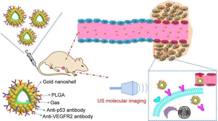
Purpose: To prospectively evaluate ultrasound molecular imaging with dual-targeted gold nanoshelled poly(lactide-co-glycolic acid) nanocapsules carrying vascular endothelial growth factor receptor type 2 (VEGFR2) and p53 antibodies (DNCs) in MCF-7 orthotopic mice model.

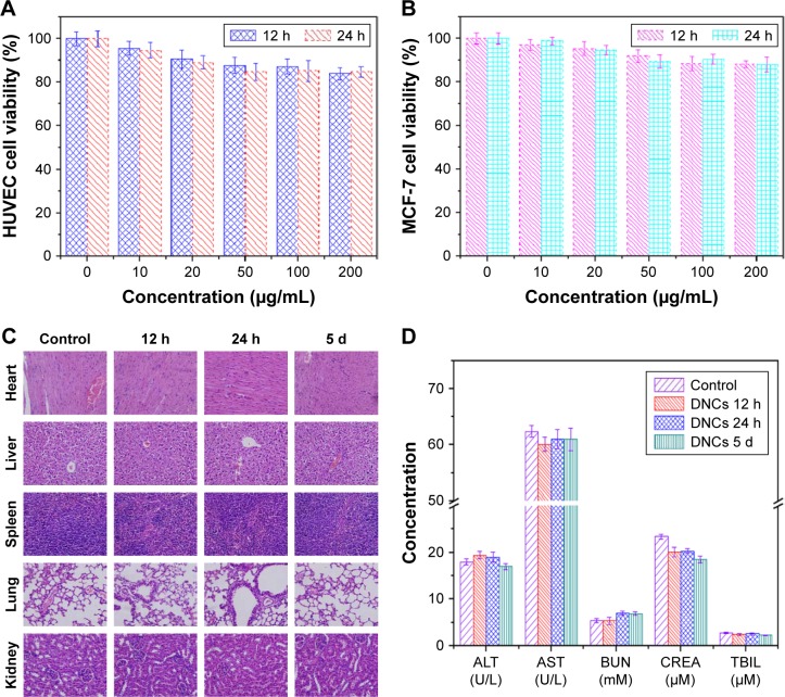
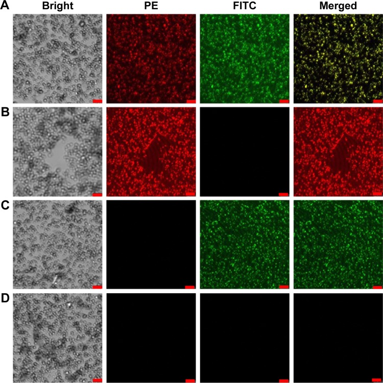
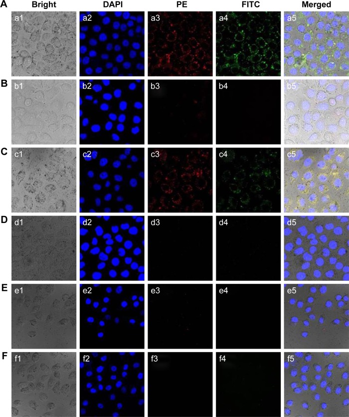
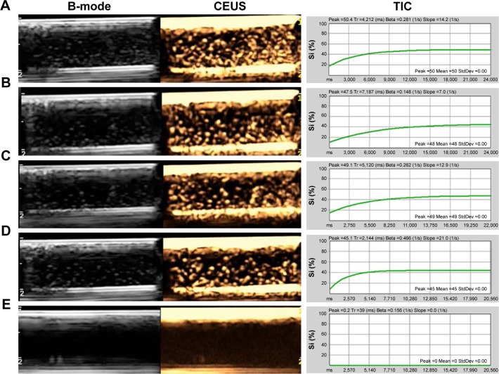
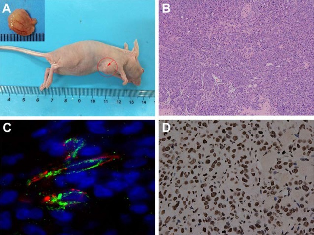
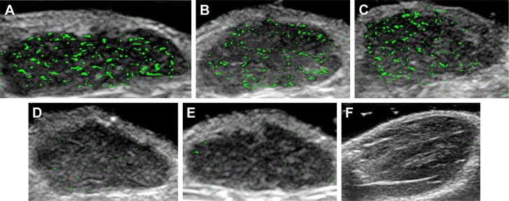
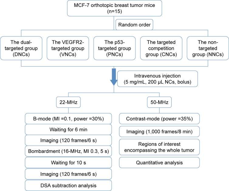
Methods: DNCs were fabricated with an inner PLGA and outer gold nanoshell spherical structure. Its targeting capabilities were evaluated by confocal laser scanning microscopy (CLSM) and flow cytometry (FCM) in vitro. Contrast-enhanced ultrasound imaging (CEUS) with DNCs was evaluated qualitatively and quantitatively in vitro and in MCF-7 orthotopic mice model by two different systems. The biodistribution of NCs in mice was preliminary investigated. Differences were calculated by using analysis of variance.

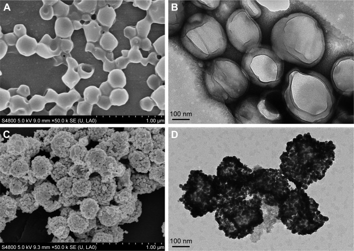
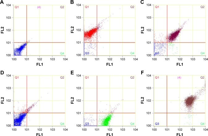
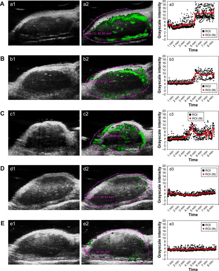
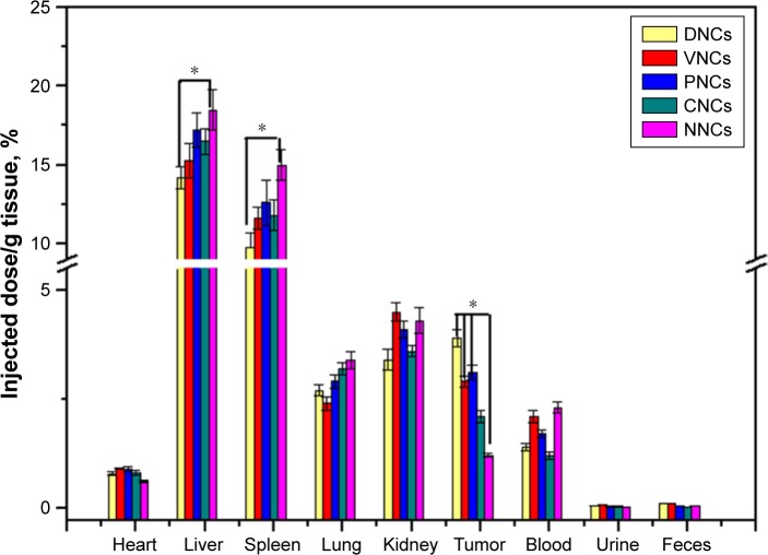
Results: DNCs showed a well-defined spherical morphology with an average diameter of 276.90±110.50 nm. In vitro, DNCs exhibited high target specificities (79.01±5.63% vs. 2.11±1.07%, P<0.01; 75.54±6.58% vs. 5.21±3.12%, P<0.01) in VEGFR2- and p53-positive cells compared with control cells. In vivo, CEUS displayed a significantly higher video intensity in two systems using DNCs in comparison with non-targeted PLGA@Au NCs and single-targeted NCs. Biodistribution studies revealed that more DNCs in breast cancer tissue could be detected in mice than in other NCs (P<0.05).

Conclusion: DNCs were demonstrated to be novel dual-targeted UCAs and may have potential applications in early non-invasive visualization of breast cancer.

Keywords: antibody; breast cancer; poly(lactic-co-glycolic acid); targeted ultrasound contrast agent; ultrasound molecular imaging.


Conflict of interest statement

Disclosure The authors report no conflicts of interest in this work.

Figures

Figure 1
Figure 1
SEM (scale =1 µm) and TEM (scale =100 nm) of PLGA NCs (A, B) and DNCs (C, D). Note: DNCs, dual-targeted gold nanoshelled poly(lactic-co-glycolic acid) nanocapsules carrying anti-vascular endothelial growth factor receptor type 2 antibody and anti-p53 antibody. Abbreviations: PLGA NCs, poly(lactic-co-glycolic acid) nanocapsules; SEM, scanning electron microscope; TEM, transmission electron microscope.
Figure 2
Figure 2
Cell viabilities of (A) HUVEC and (B) MCF-7 cells at different dosages of the DNCs (0, 10, 20, 50, 100, 200 µg/mL). (C) H&E images of heart, liver, spleen, lung, and kidney at 12 h, 24 h, and 5 days after intravenous injection of DNCs (magnification: ×100). (D) Serum biochemistry data for mice injected with saline solution and with DNCs (12 h, 24 h, and 5 days post-injection). Notes: DNCs, dual-targeted gold nanoshelled poly(lactic-co-glycolic acid) nanocapsules carrying anti-vascular endothelial growth factor receptor type 2 antibody and anti-p53 antibody. Concentration units of measure in (D) were (U/L, mM, µM). Abbreviations: ALT, alanine aminotransferase; AST, aspartate aminotransferase; BUN, blood urea nitrogen; CREA, creatinine; HUVEC, human umbilical vein endothelial cells; H&E, hematoxylin and eosin; TBIL, total bilirubin.
Figure 3
Figure 3
Confocal microscopic images of (A) DNCs, (B) VNCs, (C) PNCs, and (D) NNCs in bright field, FITC, PE, and merged channels (scale =2.5 µm), and the merged yellow signals indicate the presence of VEGFR2 and p53 in DNCs. Notes: DNCs, dual-targeted gold nanoshelled poly(lactic-co-glycolic acid) nanocapsules carrying anti-vascular endothelial growth factor receptor type 2 antibody and anti-p53 antibody; NNCs, non-targeted gold nanoshelled poly(lactic-co-glycolic acid) nanocapsules; PNCs, single-targeted gold nanoshelled poly(lactic-co-glycolic acid) nanocapsules carrying anti-p53 antibody; VNCs, single-targeted gold nanoshelled poly(lactic-co-glycolic acid) nanocapsules carrying anti-vascular endothelial growth factor receptor type 2 antibody. Abbreviations: FITC, fluorescein isothiocyanate; PE, phycoerythrin;
Figure 4
Figure 4
Confocal microscopic images of (A) HUVEC, (B) 4T1, (C) MCF-7, and (D) MDA-MB-231cells incubated with DNCs, (E) free VEGFR2-treated HUVEC cells incubated with DNCs and (F) free p53-treated HUVEC cells incubated with DNCs. Notes: DNCs, dual-targeted gold nanoshelled poly(lactic-co-glycolic acid) nanocapsules carrying anti-vascular endothelial growth factor receptor type 2 antibody and anti-p53 antibody. a1-fi: the images of bright field; a2–f2: the images of DAPI channel; a3–f3: the images of PE channel; a4–f4: the images of FITC channel; a5–f5: the images of merged channel. Abbreviations: DAPI, 4,6-diamino-2-phenyl indole; FITC, fluorescein isothiocyanate; HUVEC, human umbilical vein endothelial cells; PE, phycoerythrin; VEGFR2, vascular endothelial growth factor receptor type 2.
Figure 5
Figure 5
(A) The flow cytometry results of 4T1 incubated with DNCs. HUVEC cells incubated with VNCs and DNCs were shown in (B and C). (D) showed the results of MDA-MB-231 incubated with DNCs. MCF-7 cells incubated with PNCs and DNCs were shown in (E and F). Notes: FLI channel showed FITC signal and FL2 channel showed PE signal. DNCs, dual-targeted gold nanoshelled poly(lactic-co-glycolic acid) nanocapsules carrying anti-vascular endothelial growth factor receptor type 2 antibody and anti-p53 antibody; PNCs, single-targeted gold nanoshelled poly(lactic-co-glycolic acid) nanocapsules carrying anti-p53 antibody; VNCs, single-targeted gold nanoshelled poly(lactic-co-glycolic acid) nanocapsules carrying anti-vascular endothelial growth factor receptor type 2 antibody. Abbreviations: FITC, fluorescein isothiocyanate; HUVEC, human umbilical vein endothelial cells.
Figure 6
Figure 6
In vitro B-mode, CEUS images and TIC of (A) DNCs, (B) VNCs, (C) PNCs, (D) NNCs, and (E) pure degassed deionized water under the same concentration (0.5 mg/mL). Notes: DNCs, dual-targeted gold nanoshelled poly(lactic-co-glycolic acid) nanocapsules carrying anti-vascular endothelial growth factor receptor type 2 antibody and anti-p53 antibody; NNCs, non-targeted gold nanoshelled poly(lactic-co-glycolic acid) nanocapsules; PNCs, single-targeted gold nanoshelled poly(lactic-co-glycolic acid) nanocapsules carrying anti-p53 antibody; TIC, time-intensity curves; VNCs, single-targeted gold nanoshelled poly(lactic-co-glycolic acid) nanocapsules carrying anti-vascular endothelial growth factor receptor type 2 antibody. Abbreviations: B-mode, brightness mode; CEUS, contrast-enhanced ultrasound.
Figure 7
Figure 7
(A) MCF-7 orthotopic breast tumor nude mice model; (B) the H&E staining of tumor tissue (magnification: ×100); (C) merged confocal microscopic images of MCF-7 orthotopic breast tumor tissue after immunofluorescence staining of VEGFR2 (green), which overlap well with CD31-staining of vascular endothelial cells (red); (D) the IHC images of p53 protein (brown granules; magnification: ×400). Abbreviations: CD-31, cluster of differentiation 31; IHC, immunohistochemistry; VEGFR2, vascular endothelial growth factor receptor type 2.
Figure 8
Figure 8
Transverse color-coded US images with (A) DNCs, (B) VNCs, (C) PNCs, (D)CNCs, (E) NNCs and in (F) the hind limb adductor muscle of mice using 22-MHz probe of MCF-7 orthotopic tumor from nude mice. Notes: CNCs, free antibodies pre-treated before DNCs application; DNCs, dual-targeted gold nanoshelled poly(lactic-co-glycolic acid) nanocapsules carrying anti-vascular endothelial growth factor receptor type 2 antibody and anti-p53 antibody; NNCs, non-targeted gold nanoshelled poly(lactic-co-glycolic acid) nanocapsules; PNCs, single-targeted gold nanoshelled poly(lactic-co-glycolic acid) nanocapsules carrying anti-p53 antibody; VNCs, single-targeted gold nanoshelled poly(lactic-co-glycolic acid) nanocapsules carrying anti-vascular endothelial growth factor receptor type 2 antibody.
Figure 9
Figure 9
In vivo US imaging data using 50-MHz probe of MCF-7 orthotopic tumor from nude mice pre (a1–e1) and after (a2–e2) intravenous injection of DNCs (A), VNCs (B), PNCs (C), CNCs (D) and NNCs (E); (a3–e3) show the time-intensity curves. Notes: CNCs, free antibodies pre-treated before DNCs application; DNCs, dual-targeted gold nanoshelled poly(lactic-co-glycolic acid) nanocapsules carrying anti-vascular endothelial growth factor receptor type 2 antibody and anti-p53 antibody; NNCs, non-targeted gold nanoshelled poly(lactic-co-glycolic acid) nanocapsules; PNCs, single-targeted gold nanoshelled poly(lactic-co-glycolic acid) nanocapsules carrying anti-p53 antibody; VNCs, single-targeted gold nanoshelled poly(lactic-co-glycolic acid) nanocapsules carrying anti-vascular endothelial growth factor receptor type 2 antibody; ROI (the region of interest), the red oval region in a2-e2.
Figure 10
Figure 10
Biodistribution of Au determined by ICP-AES. Notes: CNCs, free antibodies pre-treated before DNCs application; DNCs, dual-targeted gold nanoshelled poly(lactic-co-glycolic acid) nanocapsules carrying anti-vascular endothelial growth factor receptor type 2 antibody and anti-p53 antibody; NNCs, non-targeted gold nanoshelled poly(lactic-co-glycolic acid) nanocapsules; PNCs, single-targeted gold nanoshelled poly(lactic-co-glycolic acid) nanocapsules carrying anti-p53 antibody; VNCs, single-targeted gold nanoshelled poly(lactic-co-glycolic acid) nanocapsules carrying anti-vascular endothelial growth factor receptor type 2 antibody. *Significantly different from VNCs and NNCs. Abbreviations: PLGA, poly(lactic-co-glycolic acid); ICP-AES, inductively coupled plasma atomic emission spectroscopy.
Scheme 1
Scheme 1
Schematic illustration of the structure of DNCs and the ultrasound molecular imaging process using the dual targeted agent. Note: DNCs, dual-targeted gold nanoshelled poly(lactic-co-glycolic acid) nanocapsules carrying anti-vascular endothelial growth factor receptor type 2 antibody and anti-p53 antibody. Abbreviations: PLGA, poly(lactic-co-glycolic acid); US, ultrasound; VEGFR2, vascular endothelial growth factor receptor type 2.
Scheme 2
Scheme 2
Flowchart of the process and the procedures of ultrasound imaging in vivo. Notes: CNCs, free antibodies pre-treated before DNCs application; DNCs, dual-targeted gold nanoshelled poly(lactic-co-glycolic acid) nanocapsules carrying anti-vascular endothelial growth factor receptor type 2 antibody and anti-p53 antibody; NNCs, non-targeted gold nanoshelled poly(lactic-co-glycolic acid) nanocapsules; PNCs, single-targeted gold nanoshelled poly(lactic-co-glycolic acid) nanocapsules carrying anti-p53 antibody; VNCs, single-targeted gold nanoshelled poly(lactic-co-glycolic acid) nanocapsules carrying anti-vascular endothelial growth factor receptor type 2 antibody. Abbreviations: DSA, DigSubAna, a quantitative analytical software; MI, mechanical index; NCs, nanocapsules.

Similar articles

Cited by

References

    1. Siegel RL, Miller KD, Jemal A. Cancer Statistics, 2017. CA Cancer J Clin. 2017;67(1):7–30. - PubMed
    1. Ghasemi M, Nabipour I, Omrani A, Alipour Z, Assadi M. Precision medicine and molecular imaging: new targeted approaches toward cancer therapeutic and diagnosis. Am J Nucl Med Mol Imaging. 2016;6(6):310–327. - PMC - PubMed
    1. Chakravarty R, Chakraborty S, Dash A. Molecular imaging of breast cancer: role of RGD Peptides. Mini Rev Med Chem. 2015;15(13):1073–1094. - PubMed
    1. Anderson CR, Hu X, Zhang H, et al. Ultrasound molecular imaging of tumor angiogenesis with an integrin targeted microbubble contrast agent. Invest Radiol. 2011;46(4):215–224. - PMC - PubMed
    1. Kiser J. Molecular imaging and its role in the management of breast cancer. Clin Obstet Gynecol. 2016;59(2):403–411. - PubMed

MeSH terms