Case Reports
. 2018 Feb;10(2):E120-E124.
doi: 10.21037/jtd.2018.01.126.

Initial experience with a 3D printed model for preoperative simulation of the Nuss procedure for pectus excavatum

Affiliations
Case Reports

Initial experience with a 3D printed model for preoperative simulation of the Nuss procedure for pectus excavatum

Naoto Matsuo et al. J Thorac Dis. 2018 Feb.

Abstract

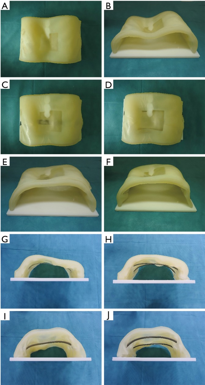
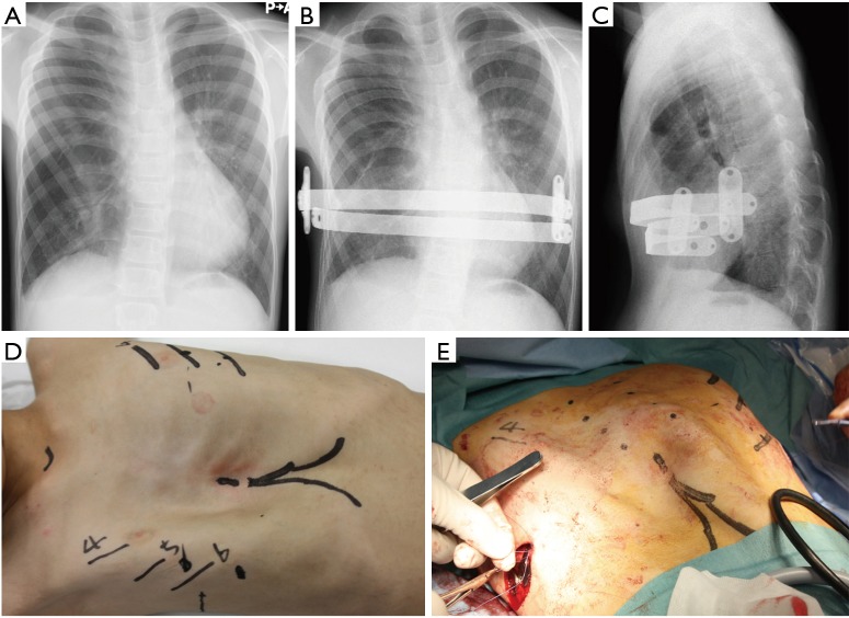
The incidence of pectus excavatum has been estimated to be between 0.1% and 0.8% though a large autopsy series reports. After publication of the Nuss procedure for pectus excavatum, it became widely accepted. However, there are still some complications, such as over-correction and recurrence. To reduce differences in the procedure due to surgeons' experience level, preoperative simulation may be useful. Thus, we performed simulated surgery using a specific patient's three-dimensional (3D) chest wall model made by a 3D printer before operation. A 13-year-old male patient with a severe deformity of the chest underwent the Nuss procedure. As in the simulation, bars were inserted into the 5th and 7th intercostal spaces (ICS), leading to improvement of the chest wall. This simulation can increase surgeons' confidence to improve the deformity by determination of the number and insertion sites of bars.

Keywords: Three-dimensional (3D) printed model; pectus excavatum; pediatric.


Conflict of interest statement

Conflicts of Interest: The authors have no conflicts of interest to declare.

Figures

Figure 1
Figure 1
Steps in making the 3D model. 3D, three-dimensional.
Figure 2
Figure 2
Simulation to insert one bar into the 6th ICS. The bar is inserted into the 6th ICS and rotated in the chest cavity (8). ICS, intercostal spaces. Available online: http://asvidett.amegroups.com/article/view/22889
Figure 3
Figure 3
Simulation to insert two bars into the 5th and 7th ICSs. Two bars are inserted into the 5th and 7th ICSs and rotated in the chest cavity (9). ICS, intercostal spaces. Available online: http://asvidett.amegroups.com/article/view/22890
Figure 4
Figure 4
Simulation of the Nuss procedure using a 3D printed model. (A) Top perspective view of chest wall before bar insertion; (B) perspective view of the chest wall from the caudal side before bar insertion; (C) top perspective view after bar insertion into the 6th ICS; (D) top perspective view after bar insertion into the 5th and 7th ICSs; (E) perspective view from the caudal side after bar insertion into the 6th ICS; (F) perspective view from the caudal side after bar insertion into the 5th and 7th ICSs; (G) perspective view from the cranial side after bar insertion into the 6th ICS; (H) perspective view from the cranial side after bar insertion into the 5th and 7th ICSs; (I) perspective view from the caudal side after bar insertion into the 6th ICS; (J) perspective view from the caudal side after bar insertion into the 5th and 7th ICSs. 3D, three-dimensional; ICS, intercostal spaces.
Figure 5
Figure 5
Improvement of the chest wall after the Nuss procedure. (A) Chest wall before the operation; (B,C) chest X-ray after the operation; (D) chest wall before the Nuss procedure; (E) chest wall after the Nuss procedure.

References

    1. Matsumoto K, Yamasaki N, Tsuchiya T, et al. Double stenting with silicone and metallic stents for malignant airway stenosis. Surg Today 2017;47:1027-35. 10.1007/s00595-016-1466-6 - DOI - PubMed
    1. Miyazaki T, Yamasaki N, Tsuchiya T, et al. Airway stent insertion simulated with a three-dimensional printed airway model. Ann Thorac Surg 2015;99:e21-3. 10.1016/j.athoracsur.2014.10.021 - DOI - PubMed
    1. Brochhausen C, Turial S, Müller FK, et al. Pectus excavatum: history, hypotheses and treatment options. Interact Cardiovasc Thorac Surg 2012;14:801-6. 10.1093/icvts/ivs045 - DOI - PMC - PubMed
    1. Croitoru DP, Kelly RE, Jr, Goretsky MJ, et al. Experience and modification update for the minimally invasive Nuss technique for pectus excavatum repair in 303 patients. J Pediatr Surg 2002;37:437-45. 10.1053/jpsu.2002.30851 - DOI - PubMed
    1. Nuss D. Minimally invasive surgical repair of pectus excavatum. Semin Pediatr Surg 2008;17:209-17. 10.1053/j.sempedsurg.2008.03.003 - DOI - PubMed

Publication types