. 2018 Jul;104(1):159-171.
doi: 10.1002/JLB.3MA1017-404R. Epub 2018 Apr 1.

DP1 receptor signaling prevents the onset of intrinsic apoptosis in eosinophils and functions as a transcriptional modulator

Affiliations

DP1 receptor signaling prevents the onset of intrinsic apoptosis in eosinophils and functions as a transcriptional modulator

Miriam Peinhaupt et al. J Leukoc Biol. 2018 Jul.

Abstract

Prostaglandin (PG) D2 is the ligand for the G-protein coupled receptors DP1 (D-type prostanoid receptor 1) and DP2 (also known as chemoattractant receptor homologous molecule, expressed on Th2 cells; CRTH2). Both, DP1 and DP2 are expressed on the cellular surface of eosinophils; although it has become quite clear that PGD2 induces eosinophil migration mainly via DP2 receptors, the role of DP1 in eosinophil responses has remained elusive. In this study, we addressed how DP1 receptor signaling complements the pro-inflammatory effects of DP2. We found that PGD2 prolongs the survival of eosinophils via a DP1 receptor-mediated mechanism that inhibits the onset of the intrinsic apoptotic cascade. The DP1 agonist BW245c prevented the activation of effector caspases in eosinophils and protected mitochondrial membranes from depolarization which-as a consequence-sustained viability of eosinophils. DP1 activation in eosinophils enhanced the expression of the anti-apoptotic gene BCL-XL , but also induced pro-inflammatory genes, such as VLA-4 and CCR3. In HEK293 cells that overexpress recombinant DP1 and/or DP2 receptors, activation of DP1, but not DP2, delayed cell death and stimulated proliferation, along with induction of serum response element (SRE), a regulator of anti-apoptotic, early-response genes. We conclude that DP1 receptors promote the survival via SRE induction and induction of pro-inflammatory genes. Therefore, targeting DP1 receptors, along with DP2, may contribute to anti-inflammatory therapy in eosinophilic diseases.

Keywords: allergy; apoptosis; inflammation; prostanoids.


Figures

Figure 1
Figure 1
DP1 receptor activation promotes survival of eosinophils. Isolated eosinophils were cultured with or without 1 μM of PGD2, DK‐PGD2, BW245c, or IL‐5 [100 pM] for 18 h. BW245c, PGD2, and IL‐5 significantly enhanced the portion of annexin V/PI eosinophils (A) and PGD2, BW245c, DK‐PGD2, and IL‐5 decreased the annexin V+ population (B) as compared to vehicle‐treated cells. (C) Shows the percentage of annexin V/PI, annexin V+/PI, annexin V+/PI+, annexin V/PI+ populations of total eosinophils at 18 h. Data show mean ± sem of 5 individual experiments using eosinophils from different donors
Figure 2
Figure 2
Activation of the DP1 receptor inhibits the intrinsic apoptotic pathway in eosinophils. Caspase 3/7 activity in eosinophils was assessed in a luminescent assay after 18 h incubation with vehicle, 1 μM of PGD2, BW245c, DK‐PGD2, or 15d‐PGJ2, Rosiglitazone (Rosi), or 100 pM of IL‐5. Cells were pretreated with 1 μM of vehicle, Cay10471 or T0070907 or increasing concentrations of MK0524 prior to agonist treatment. (A) BW245c protected from effector caspase 3/7 activity in eosinophils cultured for 18 h in serum‐reduced media (RPMI 1% FBS, 1% PenStrep). Data show means ± SEM of 5–6 individual experiments. (B) DP1 receptor antagonist MK0524 reversed the BW245c‐induced protection from caspase 3/7 activation. Data show mean ± sem of 9 individual experiments. (C) Blockade of DP2 (Cay10471) unmasks PGD2 as a potent inhibitor of caspase 3/7 activation. Data show mean ± sem of 4 individual experiments. (D) 15dPGJ2, but not rosiglitazone (Rosi), increased caspase 3/7 activity, which was not altered by the PPAR‐α antagonist T0070907 (n = 7)
Figure 3
Figure 3
Activation of the DP1 receptor maintains mitochondrial function in aging eosinophils. (A, B) Flow cytometric dot plot analysis of eosinophils stained with JC1 after 18 h incubation with 1 μM of vehicle, PGD2, BW245c, DK‐PGD2, or 50 pM of IL‐5. Depolarized mitochondrial membrane potential (MMP) is shown by the loss in yellow fluorescence (FL2). (A) Shows representative dot plots and (B) means of n = 3–4 individual experiments ± sem. **P ≤ 0.01. (C) MitoTracker Red CMXRos staining of eosinophils incubated with 1 μM of vehicle, PGD2, BW245c, and DK‐PGD2, and 100 pM of IL‐5 up to 18 h. Representative images of 3 independent experiments are shown
Figure 4
Figure 4
Bcl‐2 family protein inhibitor BH3I‐1 reverses the anti‐apoptotic effect of DP1 receptor activation. Eosinophils isolated from peripheral blood were incubated with BW245c [1 μM] in combination with or without Bcl‐XL inhibitors (A and C) BH3l‐1 [50 μM] for 5h or (B) HA14.1 [10 μM] for 3 h; (C) 18 h. (A) BH3l‐1 prevented the reduction of annexin V+ apoptotic cells induced by BW245c or IL‐5. (B) HA14.1 induced apoptosis of veh‐ and BW245c‐treated eosinophils. (C) In contrast to BH3I‐1, HA14.1 reversed the BW245c‐induced inhibition of caspase 3/7 activity. Data show mean ± sem of 5 individual experiments
Figure 5
Figure 5
PGD2 enhances the viability of HEK‐DP2+DP1 and HEK‐DP1 but not of HEK‐DP2 cells. (A) HEK293 cell lines were starved in OptiMEM for 4 h and incubated with increasing concentrations of PGD2 for 24 of 48 h. Viability was detected by MTS assay (n = 5). (B) MK0524 but not Cay10471 antagonized the viability‐enhancing action of PGD2 on HEK‐DP2+DP1. (n = 3). Data show means of 3–5 independent experiments ± sem
Figure 6
Figure 6
PGD2 treatment causes a growth advantage of HEK‐DP2+DP1 and HEK‐DP1 but not of HEK‐DP2 cells. (A) PGD2 treatment of DP1‐expressing cells led to morphologically visible growth advantage compared to HEK‐DP2 cells. Data show representative images of 4 independent experiments. (B) Cultures of serum‐starved HEK‐DP2+DP1, HEK‐DP1, or HEK‐DP2 cells were treated with increasing concentrations of PGD2 or 10% FBS and their electrical resistance was monitored for more than 60 h on an ECIS device. Data show means of 5 independent experiments + sem
Figure 7
Figure 7
DP1 induces SRE activation. (A) PGD2 activates SRE in HEK‐DP2 + DP1 and HEK‐DP1 but not in HEK‐DP2. (B) DP1 agonist BW245c induces SRE activation in HEK‐DP2 + DP1 and HEK‐DP1. (C) DP2 agonist DK‐PGD2 does not induce SRE activation. (D) Antagonists of DP1 (BWA868c) and DP2 (Cay10471) block PGD2‐ and (E) BW245c induced SRE activation in HEK‐DP2 + DP1. Data are shown as mean ± sem of 3–6 independent experiments
Figure 8
Figure 8
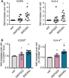
DP1 receptor agonist BW245c enhances (A) mRNA expression of CCR3 and VLA‐4 and (B) upregulates surface expression of CCR3 and VLA‐4. Isolated eosinophils (5 × 106/ml) were incubated with vehicle (EtOH), DK‐PGD2 [1 μM], or BW245c [1 μM] for 3 h (A) or 18h (B) (RPMI, 1% FBS, 1%, PenStrep, 37°C). mRNA expression was measures by qRT PCR (A). Surface expression was determined by flow cytometric analysis (B). Data show mean ± sem (n = 5–10)

Similar articles

Cited by

References

    1. Modena BD, Dazy K, White AA. Emerging concepts: mast cell involvement in allergic diseases. Transl Res. 2016;174:98–121. - PubMed
    1. Kagalwalla AF, Akhtar N, Woodruff SA, et al. Eosinophilic esophagitis: epithelial mesenchymal transition contributes to esophageal remodeling and reverses with treatment. J Allergy Clin Immunol. 2012;129:1387–1396. e7. - PMC - PubMed
    1. Yasukawa A, Hosoki K, Toda M, et al. Eosinophils promote epithelial to mesenchymal transition of bronchial epithelial cells. PLoS One. 2013;8:e64281. - PMC - PubMed
    1. Aceves SS, Broide DH. Airway fibrosis and angiogenesis due to eosinophil trafficking in chronic asthma. Curr Mol Med. 2008;8:350–358. - PubMed
    1. Wilson SJ, Rigden HM, A WardJ, et al. The relationship between eosinophilia and airway remodelling in mild asthma. Cea. 2013;43:1342–1350. - PMC - PubMed

Publication types

Substances