Crystal structure of the trimeric N-terminal domain of ciliate Euplotes octocarinatus centrin binding with calcium ions
- PMID: 29607555
- PMCID: PMC5980440
- DOI: 10.1002/pro.3418
Crystal structure of the trimeric N-terminal domain of ciliate Euplotes octocarinatus centrin binding with calcium ions
Abstract
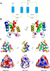
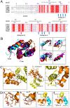
Centrin is a member of the EF-hand superfamily of calcium-binding proteins, a highly conserved eukaryotic protein that binds to Ca2+ . Its self-assembly plays a causative role in the fiber contraction that is associated with the cell division cycle and ciliogenesis. In this study, the crystal structure of N-terminal domain of ciliate Euplotes octocarinatus centrin (N-EoCen) was determined by using the selenomethionine single-wavelength anomalous dispersion method. The protein molecules formed homotrimers. Every protomer had two putative Ca2+ ion-binding sites I and II, protomer A, and C bound one Ca2+ ion, while protomer B bound two Ca2+ ions. A novel binding site III was observed and the Ca2+ ion was located at the center of the homotrimer. Several hydrogen bonds, electrostatic, and hydrophobic interactions between the protomers contributed to the formation of the oligomer. Structural studies provided insight into the foundation for centrin aggregation and the roles of calcium ions.
Keywords: aggregation; calcium coordination; centrin; crystal structure; oligomer.
© 2018 The Protein Society.
Figures


Similar articles
-
Modulation effect of double strand DNA on the self-assembly of N-terminal domain of Euplotes octocarinatus centrin.J Inorg Biochem. 2018 Mar;180:15-25. doi: 10.1016/j.jinorgbio.2017.12.001. Epub 2017 Dec 5. J Inorg Biochem. 2018. PMID: 29223826
-
Role of four conserved aspartic acid residues of EF-loops in the metal ion binding and in the self-assembly of ciliate Euplotes octocarinatus centrin.Biometals. 2016 Dec;29(6):1047-1058. doi: 10.1007/s10534-016-9975-8. Epub 2016 Oct 14. Biometals. 2016. PMID: 27743149
-
Lutetium(III)-dependent self-assembly study of ciliate Euplotes octocarinatus centrin.J Inorg Biochem. 2008 Feb;102(2):268-77. doi: 10.1016/j.jinorgbio.2007.08.010. Epub 2007 Sep 7. J Inorg Biochem. 2008. PMID: 17935787
-
Insights into functional aspects of centrins from the structure of N-terminally extended mouse centrin 1.Vision Res. 2006 Dec;46(27):4568-74. doi: 10.1016/j.visres.2006.07.034. Epub 2006 Oct 6. Vision Res. 2006. PMID: 17027898 Review.
-
Structural Basis for the Functional Diversity of Centrins: A Focus on Calcium Sensing Properties and Target Recognition.Int J Mol Sci. 2021 Nov 10;22(22):12173. doi: 10.3390/ijms222212173. Int J Mol Sci. 2021. PMID: 34830049 Free PMC article. Review.
Cited by
-
The interplay of self-assembly and target binding in centrin 1 from Toxoplasma gondii.Biochem J. 2021 Jul 16;478(13):2571-2587. doi: 10.1042/BCJ20210295. Biochem J. 2021. PMID: 34114596 Free PMC article.
References
-
- Errabolu R, Sanders MA, Salisbury JL (1994) Cloning of a cDNA encoding human centrin, an EF‐hand protein of centrosomes and mitotic spindle poles. J Cell Sci 107:9–16. - PubMed
-
- Paoletti A, Moudjou M, Paintrand M, Salisbury JL, Bornens M (1996) Most of centrin in animal cells is not centrosome‐associated and centrosomal centrin is confined to the distal lumen of centrioles. J Cell Sci 109:3089–3102. - PubMed
Publication types
MeSH terms
Substances
LinkOut - more resources
Full Text Sources
Other Literature Sources
Miscellaneous

