. 2018 Aug 15;57(16):2301-2313.
doi: 10.2169/internalmedicine.0225-17. Epub 2018 Mar 30.

Clinical Characteristics, Phenotype of Lipodystrophy and a Genetic Analysis of Six Diabetic Japanese Women with Familial Partial Lipodystrophy in a Diabetic Outpatient Clinic

Affiliations

Clinical Characteristics, Phenotype of Lipodystrophy and a Genetic Analysis of Six Diabetic Japanese Women with Familial Partial Lipodystrophy in a Diabetic Outpatient Clinic

Masanori Iwanishi et al. Intern Med. .

Abstract

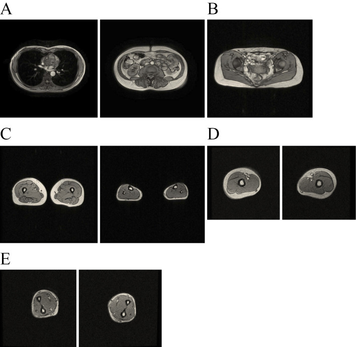
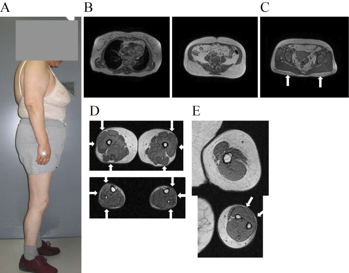
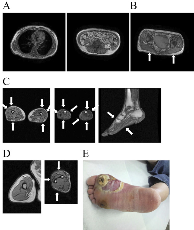
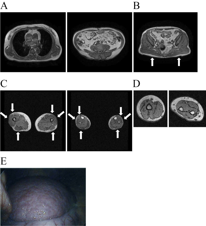
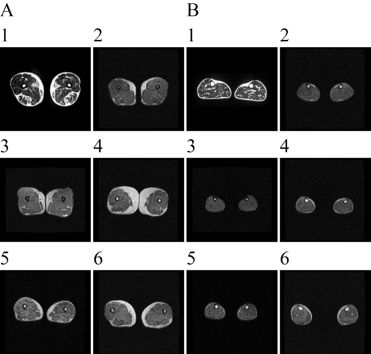
Objective Our aim was to examine the clinical characteristics and phenotype of lipodystrophy of six diabetic Japanese women with partial lipodystrophy (PL) who received a genetic analysis at a diabetic outpatient clinic. Methods We screened for PL using dual energy X-ray absorptiometry (DEXA) and magnetic resonance imaging (MRI) among patients who had a reduced peripheral skinfold thickness at the diabetic outpatient clinic of Kusatsu General Hospital between August 2003 and August 2013. We performed a mutation analysis of candidate genes, including LMNA and PPARG, in two patients with PL and whole-exome sequencing in four patients with PL. Results We identified 15 patients with PL and performed a genetic analysis in 6 of them. They had no mutations in candidate genes known to be associated with familial partial lipodystrophy (FPLD). They all had near-complete loss of subcutaneous fat, particularly in the antero-lateral and posterior thigh region and the calf region. As almost all patients were characterized by fat loss in the lower limbs with abdominal fat accumulation, a high rate of positivity for a family history, diabetes, and an unknown genetic cause, we suspected they might have FPLD1. Some patients have shown relatively severe insulin resistance, while others have shown insulin deficiency. Four and one had severe atherosclerosis and liver cirrhosis, probably due to nonalcoholic steatohepatitis, respectively. Conclusion Almost all patients with PL identified in a diabetic outpatient clinic had subcutaneous fat loss in the lower limbs with excess truncal fat and might have had FPLD1.

Keywords: diabetic outpatient clinic; familial partial lipodystrophy (FPLD); partial lipodystrophy (PL).


Figures

Figure 1.
Figure 1.
The (healthy) control subject was a 54-year-old woman. Her height was 156.0 cm, and she weighed 54.5 kg with a BMI of 22.4 kg/m2. A: Thoracic MRI scans at the level of the seventh thoracic vertebrae (left panel) and abdominal MRI scans at the umbilical level (right panel) are shown. B: T1-weighted MRI scans at the middle level of the gluteus are shown. C: MRI scans at the middle level of the thigh (left panel) and calf (right panel) are shown. She had preserved subcutaneous fat in the entire circumference of the thigh and the calf. D: MRI scans at the middle level of the right arm (left panel) and left arm (right panel) are shown. E: MRI scans at the middle level of the right forearm (left panel) and left forearm (right panel) are shown. She had preserved subcutaneous fat in the entire circumference of the arm and the forearm.
Figure 2.
Figure 2.
Patient 4 was a 69-year-old woman with a BMI of 25.5 kg/m2. A: The phenotypical features of Patient 4 are shown in the left panel. She had loss of subcutaneous fat deposits in the forearm, lower limbs, and buttocks, with prominent lower limb musculature. She had excess fat deposition around the face, neck, and trunk, although that around the face and neck has been covered with gray paper for privacy. B: Thoracic MRI scans at the level of the seventh thoracic vertebrae (left panel) and abdominal MRI scans at the umbilical level (right panel) are shown. Thoracic and abdominal MRI revealed the preservation of subcutaneous fat in the thoracic and abdominal regions of the patient. C: T1-weighted MRI scans at the middle level of the gluteus are shown. MRI scans at the middle level of the gluteus in the proband revealed a decreased amount of gluteal subcutaneous fat, indicated by arrows, compared with the subcutaneous fat in the abdominal wall, indicating mild fat loss in the buttocks. She had a decreased amount of gluteal subcutaneous fat compared with control subjects with a mean BMI of 22.4 kg/m2, as shown in Fig. 1. D: MRI scans at the middle level of the thigh (upper panel) and calf (lower panel) in the patient are shown. The patient had marked loss of subcutaneous fat, particularly in the antero-lateral and posterior thigh regions, which indicated by arrows, although subcutaneous fat in the internal thigh region remained. She had near-complete loss of subcutaneous fat in the antero-lateral and posterior calf regions, indicated by arrows, although very little subcutaneous fat in the internal calf region remained. E: MRI scans at the middle level of the left arm (upper panel) and forearm (lower panel) are shown. Axial MRI at the level of the arm revealed the preservation of subcutaneous fat. Axial MRI at the level of the forearm revealed almost complete absence of subcutaneous fat in the antero-lateral forearm, indicated by arrows, although the preservation of subcutaneous fat in the internal forearm was observed.
Figure 3.
Figure 3.
Patient 5 was a 50-year-old woman with a BMI of 22.2 kg/m2. When she came to our hospital for the treatment of a diabetic ulcer (Fig. 4E), she was found to have diabetes. A: Thoracic MRI scans at the level of the seventh thoracic vertebrae (left panel) and abdominal MRI scans at the umbilical level (right panel) are shown. Thoracic and abdominal MRI revealed the preservation of subcutaneous fat in the thoracic and abdominal regions of Patient 5. B: T1-weighted MRI scans at the middle level of the gluteus are shown. MRI scans at the middle level of the gluteus in the proband revealed a decreased amount of gluteal subcutaneous fat, indicated by arrows, compared with control subjects with a BMI of 22.4 kg/m2, as shown in Fig. 1. C: MRI scans at the middle level of the thigh (left panel) and calf (middle panel) in the patient are shown, as are scans at the level of the right leg (right panel). The patient had very little subcutaneous fat, particularly in the antero-lateral and posterior thigh regions, indicated by arrows, although subcutaneous fat in the internal thigh region remained. She had near-complete loss of subcutaneous fat in the entire circumference of the calf, indicated by arrows. MRI scans of right leg revealed almost complete absence of dorsal and plantar subcutaneous fat, indicated by arrows. D: MRI scans at the middle level of the right arm (left panel) and forearm (right panel) are shown. Axial MRI at the middle level of the arm revealed the preservation of subcutaneous fat. Axial MRI at the level of the forearm revealed almost complete absence of subcutaneous fat in the antero-lateral and posterior forearm regions, indicated by arrows, although small amounts of subcutaneous fat in the internal forearm were preserved. E: The diabetic ulcer at her first visit is shown.
Figure 4.
Figure 4.
Patient 6 was a 67-year-old woman with a BMI of 25.2 kg/m2. A: Thoracic MRI scans at the level of the sixth thoracic vertebrae (left panel) and abdominal MRI scans at the umbilical level (right panel) are shown. Thoracic and abdominal MRI revealed the preservation of subcutaneous fat in the thoracic and abdominal regions of Patient 6. B: T1-weighted MRI scans at the middle level of the gluteus are shown. MRI scans at the middle level of the gluteus in the proband revealed a decreased amount of gluteal subcutaneous fat, indicated by arrows, compared with control subjects with a mean BMI of 22.4 kg/m2, as shown in Fig. 1. C: MRI scans at the middle level of the thigh (left panel) and calf (right panel) in the patient are shown. The patient had marked loss of subcutaneous fat, particularly in the antero-lateral and posterior thigh regions, indicated by arrows, although subcutaneous fat in the internal thigh region remained. She had marked loss of subcutaneous fat in the antero-lateral and posterior calf regions, indicated by arrows, although little subcutaneous fat in the internal calf region remained. D: MRI scans at the middle level of the right arm (left panel) and forearm (right panel) are shown. Axial MRI at the middle level of the arm revealed the preservation of subcutaneous fat, as did that at the middle level of the forearm. E: Nodular formation on the surface of the enlarged left hepatic lobe, consistent with liver cirrhosis or pre-cirrhosis, was shown by peritoneoscopy when Patient 6 underwent laparoscopic cholecystectomy for cholelithiasis at 67 years of age.
Figure 5.
Figure 5.
A: MRI scans at the middle level of the thigh in six patients are shown. All patients had near-complete loss of subcutaneous fat in the antero-lateral and posterior thigh region, although the amounts of subcutaneous fat in the internal thigh region were preserved in these patients. B: MRI scans at the middle level of the calf in six patients are shown. Patient 1 had very little subcutaneous fat in the entire circumference of the calf, while Patients 2, 3, and 5 had near-complete loss of subcutaneous fat in the entire circumference of the calf. Patients 4 and 6 had near-complete loss of subcutaneous fat in the calf region, particularly in the antero-lateral and posterior calf regions, although they had little subcutaneous fat in the internal calf region.

Similar articles

Cited by

References

    1. Hussain I, Garg A. Lipodystrophy syndromes endocrinol. Metab Clin N Am 45: 783-797, 2016. - PMC - PubMed
    1. Brown RJ, Araujo-Vilar D, Cheung PT, et al. . The diagnosis and management of lipodystrophy syndrome: a multi-society practice guideline. J Clin Endocrinol Metab 85: 1-14, 2016. - PMC - PubMed
    1. Agarwal AK, Garg A. Genetic basis of lipodystrophies and management of metabolic complications. Annu Rev Med 57: 297-311, 2006. - PubMed
    1. Garg A. Lipodystrophies: genetic and acquired body fat disorders. J Clin Endocrinol Metab 96: 3313-3325, 2011. - PMC - PubMed
    1. Strickland LR, Lok K, Guo F, Garvey WT. Type 2 Diabetes with partial lipodystrophy of the limbs. Diabetes Care 36: 2247-2253, 2013. - PMC - PubMed