Review
. 2018 Apr 2;128(4):1217-1228.
doi: 10.1172/JCI95146. Epub 2018 Apr 2.

Senescent cells: a therapeutic target for cardiovascular disease

Affiliations
Review

Senescent cells: a therapeutic target for cardiovascular disease

Bennett G Childs et al. J Clin Invest. .

Abstract

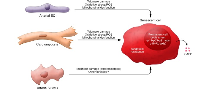
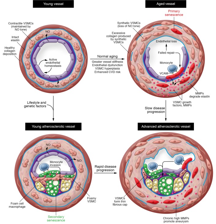
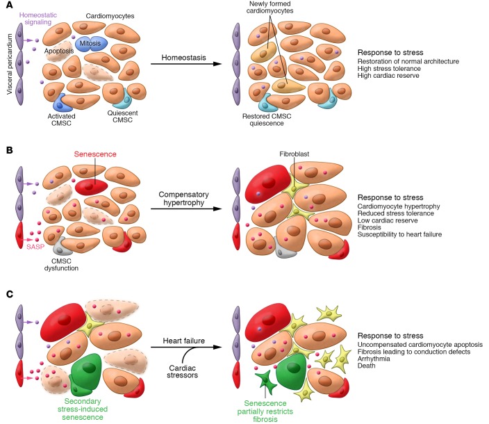
Cellular senescence, a major tumor-suppressive cell fate, has emerged from humble beginnings as an in vitro phenomenon into recognition as a fundamental mechanism of aging. In the process, senescent cells have attracted attention as a therapeutic target for age-related diseases, including cardiovascular disease (CVD), the leading cause of morbidity and mortality in the elderly. Given the aging global population and the inadequacy of current medical management, attenuating the health care burden of CVD would be transformative to clinical practice. Here, we review the evidence that cellular senescence drives CVD in a bimodal fashion by both priming the aged cardiovascular system for disease and driving established disease forward. Hence, the growing field of senotherapy (neutralizing senescent cells for therapeutic benefit) is poised to contribute to both prevention and treatment of CVD.


Conflict of interest statement

Conflict of interest: JMVD is a cofounder of Unity Biotechnology, a company developing senolytic medicines, including small molecules that selectively eliminate senescent cells. JMVD, HL, and BGC are co-inventors on patent applications licensed to or filed by Unity Biotechnology (15/455,630 and 62/057,825).

Figures

Figure 1
Figure 1. Key properties of senescent cells.
In response to various types of stressors (generally irreparable macromolecular damage), replication-competent cardiovascular cells undergo senescence. The 3 major hallmarks of SNCs are a permanent cell cycle arrest, mediated by signaling through the p19Arf-p53-p21Cip/Waf and p16Ink4a-Rb axes; apoptosis resistance, achieved by upregulation of prosurvival factors; and acquisition of the senescence-associated secretory phenotype (SASP), a bioactive secretome containing cytokines, growth factors, proteases, and other signaling molecules.
Figure 2
Figure 2. Cellular senescence is a cause and consequence of atherosclerosis.
Both primary (age-derived) SNCs and secondary (atherosclerosis-derived) SNCs may promote atherosclerosis. In young arteries (top left), senescence burden is low. Vessel homeostasis is achieved by endothelium-derived NO acting on VSMCs, promoting vasodilation, quiescence, contractile phenotype, and endothelial health. During normal aging (top right), endothelial cells and VSMCs undergo primary senescence (red cells). Loss of NO tone may arise due to failure of endothelial cell homeostasis, leading to endothelial dysfunction and senescence. Excessive VSMC proliferation in aging vessels may also drive VSMC senescence, further exacerbating VSMC hyperplasia via SASP-mediated growth factor release. Whatever the causal chain, loss of NO tone converts VSMC to the synthetic phenotype, prompting VSMC hyperplasia, medial thickening, collagen overproduction, and synthesis of elastin-degrading metalloproteases (MMPs), collectively producing vessel stiffening, widened pulse pressure, hypertension, and increased cardiac afterload. Vessel stiffening interrupts normal shear stress signals that suppress VCAM expression, with proatherogenic effects. Absence of risk factors in late life may produce late-onset cardiovascular disease (CVD; bottom right). Alternatively, early-life CVD risk factors (e.g., diabetes, hypertension, metabolic syndrome, and dyslipidemia) promote aberrant oxi-LDL accumulation in the subendothelial space, initially producing secondary senescent foam cell macrophages. At later stages of disease, secondary senescent foamy endothelium and VSMCs also arise (green cells). This faster trajectory results from intense, proatherogenic stress and results in early CVD (bottom left). The relative contribution of primary and secondary SNCs to atheroprogression probably varies with life history and personal risk factors, but both types likely contribute to key features of clinical disease, including thinning of the fibrous cap as a result of VSMC growth arrest and SASP-derived collagenases and elastases; enhanced lesion growth due to expression of VCAM and ICAM; and lesion-associated aneurysm resulting from medial elastin degradation.
Figure 3
Figure 3. Senescent cells drive heart failure.
(A) In young hearts, myocardium homeostasis, normal tissue function, and excess cardiac reserve are maintained through de novo formation of cardiomyocytes from resident cardiomyocyte stem cells (CMSCs) or division of incompletely differentiated, small cardiomyocytes. New cardiomyocyte formation matches a relatively low rate of cardiomyocyte apoptosis (purple cells). Appropriate CMSC function, including return to quiescence and suppression of cardiomyocyte death, may be maintained by homeostatic signals arising from the pericardium. (B) In disease-free, aging hearts, a declining ability to replace apoptotic cardiomyocytes due to progenitor dysfunction/senescence conspires with an increasing rate of cardiomyocyte death to produce compensatory hypertrophy. In the resting state and in the absence of further CVD, this adaptive change partially preserves heart function but predisposes to heart failure upon further stress. Interestingly, the INK-ATTAC SNC-killing transgenic strategy efficiently removes SA β-gal–positive pericardial SNCs from aged mice and prevents both cardiomyocyte hypertrophy and declining β-adrenergic stress tolerance. Based on this, we propose the existence of pericardium-derived prohomeostatic signaling that is disrupted during aging by pericardial SASP factors, although similar disruptive signaling may arise from senescent cardiomyocytes, CMSCs, or other cell types in the myocardium. (C) When challenged with cardiovascular stressors, such as ischemia in coronary artery disease, diabetes, or hypertension, the aged myocardium exceeds its functional reserve and decompensates. Apoptotic cardiomyocytes are not replaced, hypertrophy no longer preserves function, and excessive fibrosis leads to conduction defects, arrhythmia, and HF. To some extent, fibroblast senescence restricts fibrosis, but the long-term presence of these and other SNC types in the failing heart is suspected to be deleterious. It is currently unclear whether SNC removal can improve established heart failure or merely blunts development.

References

    1. Childs BG, Durik M, Baker DJ, van Deursen JM. Cellular senescence in aging and age-related disease: from mechanisms to therapy. Nat Med. 2015;21(12):1424–1435. doi: 10.1038/nm.4000. - DOI - PMC - PubMed
    1. Roger VL, et al. Heart disease and stroke statistics — 2011 update: a report from the American Heart Association. Circulation. 2011;123(4):e18–e209. doi: 10.1161/CIR.0b013e3182009701. - DOI - PMC - PubMed
    1. Feingold KR, Grunfeld C. Approach to the patient with dyslipidemia. In: De Groot G, et al., eds. Endotext [Internet]. South Dartmouth, Massachusetts, USA; 2000. https://www.ncbi.nlm.nih.gov/books/NBK278943/ Accessed January 29, 2018.
    1. Acelajado MC, et al. Refractory hypertension: definition, prevalence, and patient characteristics. J Clin Hypertens (Greenwich) 2012;14(1):7–12. doi: 10.1111/j.1751-7176.2011.00556.x. - DOI - PMC - PubMed
    1. Upadhya B, Taffet GE, Cheng CP, Kitzman DW. Heart failure with preserved ejection fraction in the elderly: scope of the problem. J Mol Cell Cardiol. 2015;83:73–87. doi: 10.1016/j.yjmcc.2015.02.025. - DOI - PMC - PubMed

Publication types