. 2018 May;7(5):1988-2002.
doi: 10.1002/cam4.1455. Epub 2018 Apr 2.

Regulation of NCAPG by miR-99a-3p (passenger strand) inhibits cancer cell aggressiveness and is involved in CRPC

Affiliations

Regulation of NCAPG by miR-99a-3p (passenger strand) inhibits cancer cell aggressiveness and is involved in CRPC

Takayuki Arai et al. Cancer Med. 2018 May.

Abstract

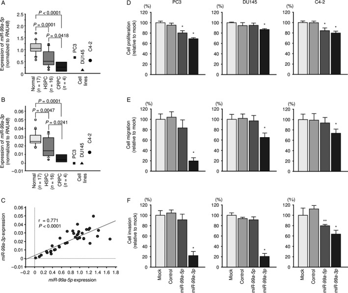
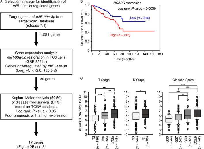
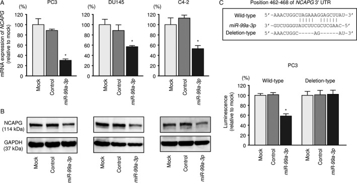
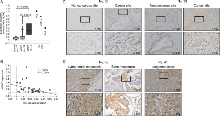
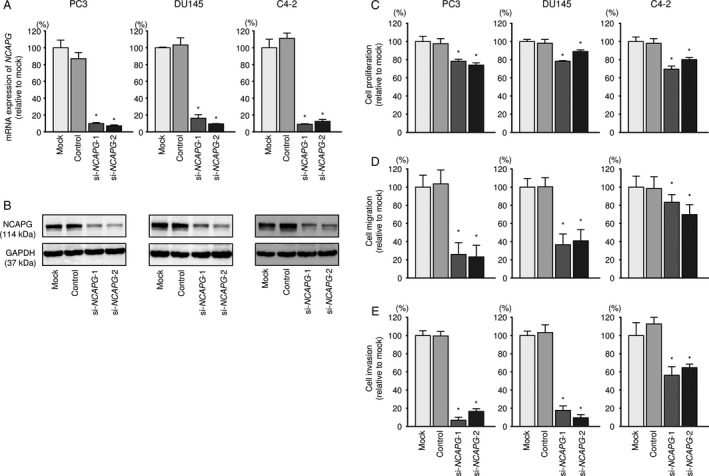
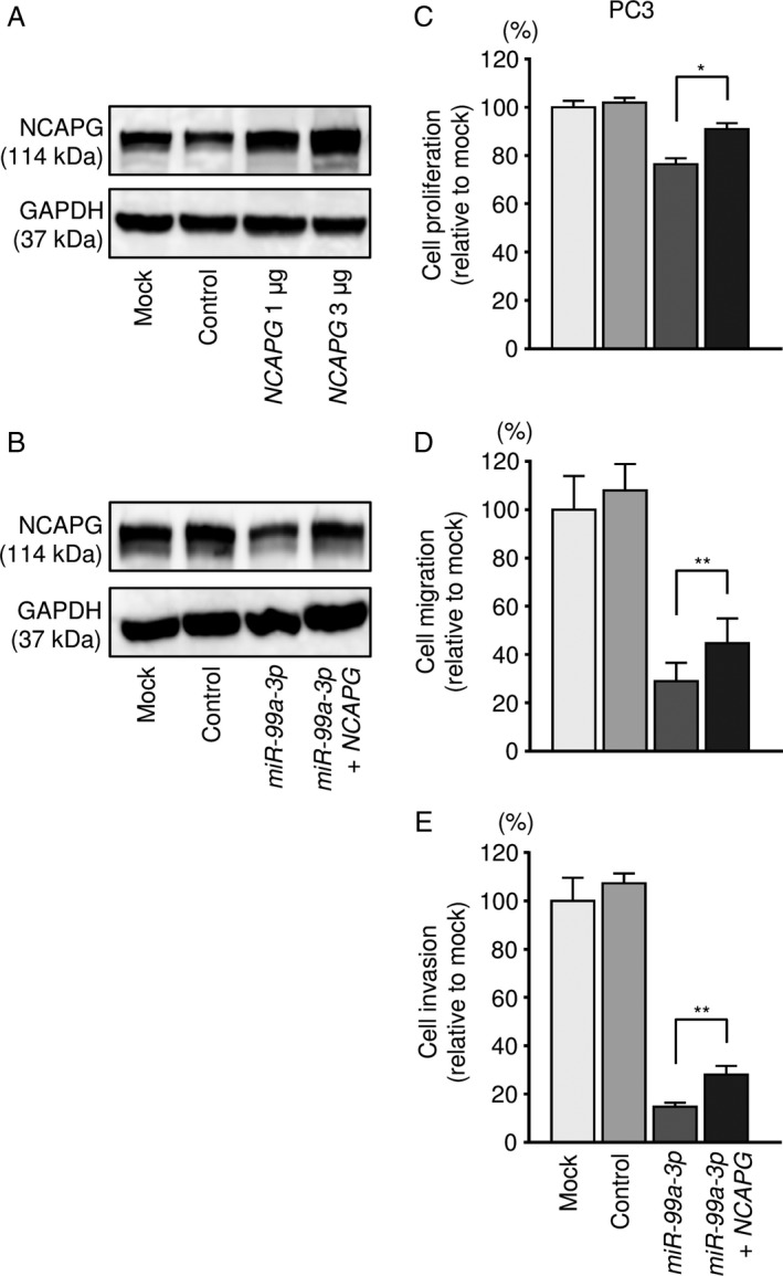
Effective treatments for patients with castration-resistant prostate cancer (CRPC) have not yet been established. Novel approaches for identification of putative therapeutic targets for CRPC are needed. Analyses of RNA sequencing of microRNA (miRNA) expression revealed that miR-99a-3p (passenger strand) is significantly downregulated in several types of cancers. Here, we aimed to identify novel miR-99a-3p regulatory networks and therapeutic targets for CRPC. Ectopic expression of miR-99a-3p significantly inhibited cancer cell proliferation, migration, and invasion in PCa cells. Non-SMC condensin I complex subunit G (NCAPG) was a direct target of miR-99a-3p in PCa cells. Overexpression of NCAPG was detected in CRPC clinical specimens and was significantly associated with shorter disease-free survival and advanced clinical stage. Knockdown of NCAPG inhibited cancer cell aggressiveness. The passenger strand miR-99a-3p acted as an antitumor miRNA in naïve PCa and CRPC. NCAPG was regulated by miR-99a-3p, and its overexpression was involved in CRPC pathogenesis. Involvement of passenger strand of miRNA in cancer pathogenesis is novel concept, and identification of antitumor miRNA regulatory networks in CRPC might be provided novel prognostic markers and therapeutic targets for this disease.

Keywords: miR-99a-3p; miR-99a-5p; Castration-resistant prostate cancer; microRNA; non-SMC condensin I complex subunit G.


Figures

Figure 1
Figure 1
Expression of miR‐99a‐5p/3p in clinical prostate specimens and functional analysis of miR‐99a‐5p/3p in PCa cell lines. (A) Expression levels of miR‐99a‐5p in PCa clinical specimens and cell lines determined using qRTPCR. RNU48 was used as an internal control. (B) Expression levels of miR‐99a‐3p in PCa clinical specimens and cell lines. (C) Correlations among the relative expression levels of miR‐99a‐5p and miR‐99a‐3p. (D‐F) Cell proliferation, migration, and invasion assays in cells transfected with miR‐99a‐5p/3p. *< 0.0001 and **< 0.001.
Figure 2
Figure 2
Identification of miR‐99a‐3p target genes and relationship between NCAPG and clinicopathological factors. (A) Flowchart of the strategy for identification of miR‐99a‐3p target genes. (B) Kaplan–Meier patient survival curves for disease‐free survival rates based on NCAPG expression in patients with PCa from TCGA database. (C) According to TCGA database, the expression levels of NCAPG were significantly increased in cases of advanced T stage, advanced N stage, and high Gleason score. *< 0.01, **< 0.001, and ***< 0.0001.
Figure 3
Figure 3
Kaplan–Meier survival curves based on expression of 16 genes, excluding NCAPG, in patients with PCa. Kaplan–Meier patient survival curves for disease‐free survival rates based on expression of 16 genes, excluding NCAPG, in patients with PCa, according to TCGA database.
Figure 4
Figure 4
Direct regulation of NCAPG by miR‐99a‐3p in PCa cells. (A) NCAPG mRNA expression was evaluated using qRTPCR in PC3, DU145, and C4‐2 cells 48 h after transfection with miR‐99a‐3p. GAPDH was used as an internal control. *< 0.0001. (B) NCAPG protein expression was evaluated by Western blotting in PC3, DU145, and C4‐2 cells 72 h after transfection with miR‐99a‐3p. (C) miR‐99a‐3p binding sites in the 3′‐UTR of NCAPG mRNA. Dual‐luciferase reporter assays in PC3 using vectors encoding a putative miR‐99a‐3p target site in the NCAPG 3′‐UTR (positions 462–468). Data were normalized by expression ratios of Renilla/firefly luciferase activities. *< 0.0001.
Figure 5
Figure 5
Expression of NCAPG in clinical PCa specimens. (A) Expression levels of NCAPG in PCa clinical specimens and cell lines. GUSB was used as an internal control. (B) The negative correlation between miR‐99a‐3p and NCAPG. (C) Immunochemical staining of NCAPG in HSPC specimens. (D) Immunochemical staining of NCAPG in mCRPC specimens.
Figure 6
Figure 6
Effects of NCAPG silencing in PCa cell lines. (A) NCAPG mRNA expression was evaluated using qRTPCR analysis of PC3, DU145, and C4‐2 cells 48 h after transfection with siNCAPG‐1 or siNCAPG‐2. GAPDH was used as an internal control. *< 0.0001. (B) NCAPG protein expression was evaluated by Western blot analysis of PC3, DU145, and C4‐2 cells 72 h after transfection with siNCAPG‐1 or siNCAPG‐2. GAPDH was used as a loading control. (C‐E) Cell proliferation, migration, and invasion assays following transfection with si‐NCAPG‐1 and si‐NCAPG‐2. *< 0.0001.
Figure 7
Figure 7
Effects of cotransfection with NCAPG/miR‐99a‐3p in PCa cell lines. (A) NCAPG protein expression was evaluated by Western blot analysis of PC3 cells 48 h after forward transfection with the NCAPG vector. GAPDH was used as a loading control. (B) NCAPG protein expression was evaluated by Western blot analysis of PC3 cells 72 h after reverse transfection with miR‐99a‐3p and 48 h after forward transfection with the NCAPG vector. (C) Cell proliferation was determined using XTT assays 72 h after reverse transfection with miR‐99a‐3p and 48 h after forward transfection with the NCAPG vector. *< 0.0001. (D) Cell migration activity was assessed by wound‐healing assays 48 h after reverse transfection with miR‐99a‐3p and 24 h after forward transfection with the NCAPG vector. **< 0.001. (E) Cell invasion activity was characterized by invasion assays 48 h after reverse transfection with miR‐99a‐3p and 24 h after forward transfection with the NCAPG vector. **< 0.001.

References

    1. Siegel, R. L. , Miller K. D., and Jemal A.. 2017. Cancer statistics, 2017. CA Cancer J. Clin. 67:7–30. - PubMed
    1. Crona, D. J. , and Whang Y. E.. 2017. Androgen receptor‐dependent and ‐independent mechanisms involved in prostate cancer therapy resistance. Cancers (Basel) 9:pii: E67. - PMC - PubMed
    1. Crawford, E. D. , Higano C. S., Shore N. D., Hussain M., and Petrylak D. P.. 2015. Treating patients with metastatic castration resistant prostate cancer: a comprehensive review of available therapies. J. Urol. 194:1537–1547. - PubMed
    1. Bartel, D. P. 2004. MicroRNAs: genomics, biogenesis, mechanism, and function. Cell 116:281–297. - PubMed
    1. Filipowicz, W. , Bhattacharyya S. N., and Sonenberg N.. 2008. Mechanisms of post‐transcriptional regulation by microRNAs: are the answers in sight? Nat. Rev. Genet. 9:102–114. - PubMed

Publication types

MeSH terms